Download
1 / 20
210 likes | 398 Views
Amino Acids – High Chemical Diversity , Low Structural Diversity.

E N D
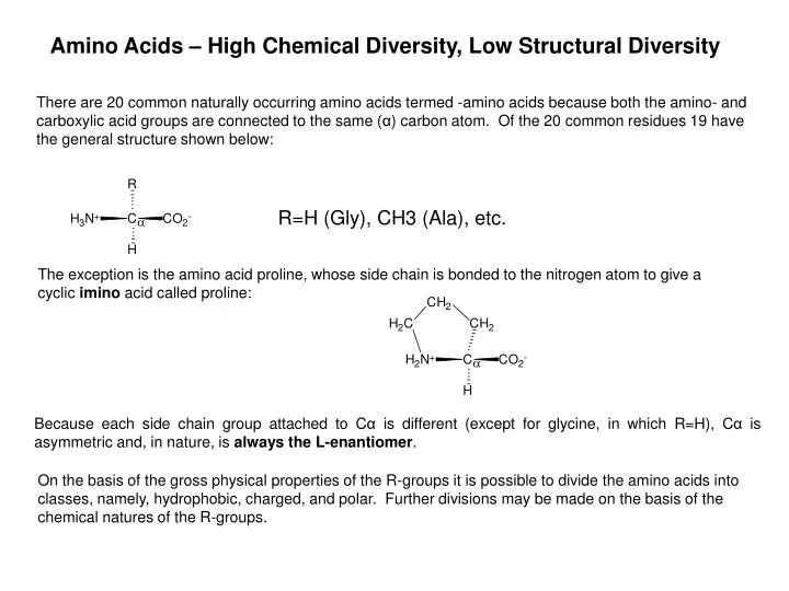
Amino Acids – High Chemical Diversity, Low Structural Diversity There are 20 common naturally occurring amino acids termed -amino acids because both the amino- and carboxylic acid groups are connected to the same (α) carbon atom. Of the 20 common residues 19 have the general structure shown below: • R=H (Gly), CH3 (Ala), etc. The exception is the amino acid proline, whose side chain is bonded to the nitrogen atom to give a cyclic imino acid called proline: Because each side chain group attached to Cα is different (except for glycine, in which R=H), Cα is asymmetric and, in nature, is always the L-enantiomer. On the basis of the gross physical properties of the R-groups it is possible to divide the amino acids into classes, namely, hydrophobic, charged, and polar. Further divisions may be made on the basis of the chemical natures of the R-groups.
Proteins: One Linkage Position and Configuration The Peptide Bond x • With few exceptions, peptide groups assume a trans-conformation, in which successive Cα atoms are on opposite sides of the peptide bond joining them. The observation that the peptide bond prefers to be planar is best explained with reference to the molecular orbitals. Maximum orbital overlap (and so maximum stabilization) is achieved when the p-type orbital of the N-atom is parallel to the p-orbitals of the π‑bond. During rotation around the C—N bond the overlap decreases and becomes nearly zero when the orbitals are perpendicular to each other. This is a higher energy conformation, because it lacks the stabilizing orbital interactions. Thus, rotation is energetically disfavoured, and the bond remains close to planarity. These characteristics are important because they indicate that the backbone of a protein is a linked sequence of rigid planar peptide groups. The protein backbone conformation can therefore be specified by the torsion (or dihedral) angles about the Cα—N bond (φ-torsion angle) and Cα—C bond (ψ-torsion angle) of each of its amino acid residues.
Carbohydrates – Huge Structural Diversity, Low Chemical Diversity
Oligosaccharides: Multiple Linkage Positions and Configurations Glc-α-(1-4)-Glc (Starch) Glc-β-(1-4)-Glc (Cellulose) The same two amino acids 1 possible peptide The same two monosaccharides 20 possible disaccharides Avian Flu Receptor Human Flu Receptor α-(2-3)-Gal versusα-(2-6)-Gal
Carbohydrate-Protein Interactions in Cell Life and Death Other Cells Hormones Viruses Toxins Cell Bacteria
Enthalpy (ΔH) and Entropy (ΔS) Electrostatic Interactions (Hydrogen-bonds, charge-charge, charge-dipole, dipole-dipole) Dispersive Interactions (Van der Waals attractions and repulsions) ΔH < 0 reaction is exothermic, tells us nothing about the spontaneity of the reaction • Δ H > 0 reaction is endothermic, tells us nothing about the spontaneity of the reaction Examples: Oxidation of glucose: C6H12O6 + 6O2 6CO2 + 6H2O ΔH = -2803 kJmol-1 • Just because a reaction is exothermic (that is because ΔH < 0) does not mean that it is spontaneous. And what about Dissolving salt: NaCl(s) + H2O(l) Na+(aq) + Cl-(aq) H = 4 kJmol-1 Just because this reaction is endothermic (ΔH > 0) does not mean that it doesn’t happen. • Enthalpy alone is not sufficient to decide whether a reaction will occur. The missing factor is called Entropy or ΔS. Entropic Contributions: Solute Related (conformational entropy) Solvent Related (ligand and receptor desolvation) • ΔS < 0 reaction leads to order, tells us nothing about the spontaneity of the reaction • ΔS > 0 reaction leads to disorder, tells us nothing about the spontaneity of the
Thermodynamics of Ligand-Protein Interactions (ΔG) • Remember: ΔGreaction = ΔGproducts – ΔGreactants • ΔG = ΔH - T ΔS, • The reaction is favourable only when ΔG < 0 Ligand Binding Energy is also computed as if it were a reaction: Ligand + Receptor → Complex • ΔGBinding= ΔGComplex – ΔGLigand – ΔGReceptor • = (ΔHComplex – T Δ SComplex) – (ΔHLigand – T ΔSLigand) – (ΔHReceptor – T ΔSReceptor) There is a temptation to draw conclusions only from the structure of the complex, but: • ΔGBinding ≠ MM EnergyComplexMM “Energy” is often just the potential energy from a force fieldcalculation. • MM Energy often ignores entropy and desolvation! • and is often NOT computed as a difference between reactants and products! • Bad modeling can’t be trusted!
Energetic Contributions to Ligand Binding Hydrogen Bonds A molecule which has a weakly acidic proton (O—H, N—H) may function as a proton donor (DH) in a hydrogen bond with another molecule in which an electronegative atom (O, N) is present to act as an acceptor (A). • D—H ···A— • A typical hydrogen bond between polar uncharged groupshas its maximum stability at an interatomic (A ···D) separation of 2.7-3.1 Å and may contribute up to approximately 5 kcalmol-1 in the gas phase. Hydrogen bonds show a high dependence on the orientation of the donor and acceptor groups, with a tendency for the D—H ··· A angle to be linear. D—H ··· ··· ··· ··· A X-ray crystallographic studies of sugar-protein complexes can provide detailed structural information pertaining to hydrogen bonding in the binding site.
Hydrogen Bonds, Continued The hydrophilic nature of sugars arises from the presence of hydroxyl groups attached typically to 5 out of 6 of the carbon atoms of the sugar: The polyhydroxylated structure of a sugar has often been cited in support of the importance of hydrogen bonding in the interaction between the sugar and either a receptor or with solvent. For example, in the case of arabinose binding protein, the arabinopyranose is involved in approximately 54 hydrogen bonds either with the protein, or with coordinated water molecules. A B A. Typical hydrogen bonds between a sugar and a protein. B. Between a sugar and water.
Effect of Loss of a Hydrogen Bond on Binding Energies The presence of hydrogen bonding is essential to the binding of a sugar to a protein. If there are not at least as many hydrogen bonds in the complex as there are between the sugar and the solvent, the binding will not be ENTHALPICALLY favored. If each hydrogen bond stabilizes the interaction by 5 kcal/mol, the loss of a single hydrogen bond would severely diminish the binding affinity. • Consider two ligands: one makes 4 hydrogen bonds to the receptor, the other makes 3 hydrogen bonds. L1 + Receptor → Complex1 GBinding (L1) -20 kcal/mol L2 + Receptor → Complex2 GBinding (L2) -15 kcal/mol G = GBinding (L1) – GBinding (L2) = -5 kcal/mol (favoring the binding of L1) How much would the loss of a single hydrogen bond change the binding affinities? Recall: G = –RT ln(K) G = –RTln(K1) – RTln(K2) = –RT(ln(K1) – ln(K2)) = –RTln(K1/K2) So for the two ligands, the ratio of their binding affinities (at 293 K) is: -5 = –RT ln(K1/K2) = –0.00198∙398ln(K1/K2) = –0.59 ln(K1/K2) Therefore K1/K2 = e8.47 = 4788, so the net loss of a hydrogen bond decreases affinity ~ 5000 fold. But why isn’t counting H-bonds a good measure of affinity?
Since many sugars all have the same number of hydroxyl groups and differ only in the configuration of the hydroxyl groups, they all can exhibit very similar hydrogen bonding patterns if they can physically fit into the receptor site. • The Role of Water Each hydroxyl group in a sugar may act as both a proton acceptor and a proton donor in hydrogen bonds. In solution it is possible for two water molecules to orient themselves along each sugar hydroxyl group lone-pair axis and so an optimum hydrogen bonding network is present. However in the complex it may not be possible to orient the protein side chains as optimally. Since for every hydrogen bond the sugar forms with the protein, it must break at least one with the water, thus the net ENTHALPIC gain from hydrogen bonding may be relatively small. Consequently, while hydrogen bonding is essential to the binding of the sugar, it is not sufficient to generate very tight binding, or to discriminate between different sugars. This may explain why monosaccharide-protein interactions are often very weak: Ka=102-3 M‑1.
Example: Effect of loss of OH on affinity Antigenic oligosaccharides from S. flexneri Y with antibody SYAJ6 • Vyas, N.K., et al., Biochemistry, 2002. 41: p. 13575-13586.
Other Enthalpic Contributions to Binding Van der Waals Interactions (instantaneous dipole - induced dipole) As any pair of atoms approach each other a weak attraction develops that is called a van der Waals interaction. In order to provide a noticeable ENTHALPIC benefit the atoms must be no further apart than the sum of their van der Waals radii (typically less than ~ 4 Å). In a carbohydrate protein complex there may be many such interactions, and although each one provides very little energy, their sum may be significant. The maximum energetic contribution from vdw interaction is small (only about 0.2 kcal/mol) per interacting atom and decreases with distance as a function of 1/r6. The combination of vdw and hydrophobic effects can be significant. For example: in the antigenic oligosaccharide from the bacterial polysaccharide of V. choleraetypes Ogawa (OMe) and Inaba (OH). Antibodies against Ogawa do not bind to Inaba! Because of the extreme sensitivity of the energies of van der Waals contacts to interatomic distance, a slight change in ligand shape or binding orientation can greatly alter the number of van der Waals contacts. Thus, carbohydrate ligand specificity depends very highly on van der Waals contacts.
Example: Effect of loss of van der Waals and hydrophobic contacts on affinity Antigenic oligosaccharides from V. cholerae with antibodyS20-4 Too few favorable interactions or too many unfavorable ones will hurt binding Ogawa Inaba • Affinities of Vibriocholerae O:1 Serotypes Ogawa and Inaba for mAb S20-4 • Wang, J., et al., J. Biol. Chem., 1998. 273(5): p. 2777-2783.
Effect of Entropy on Binding Energies A large contributor to the ENTROPY of binding is from the release of water molecules. This arises from two contributions, desolvation entropy and the hydrophobic effect. Desolvation Entropy As already seen, when the sugar binds to the protein, it displaces water molecules that were previously present in the binding site. It also must release water molecules that were directly coordinated to the sugar itself. This release of water results in an increase in the entropy of the system, i.e. SBinding > 0 and so -TSBinding < 0. But the desolvation free energy may still be unfavorable (>0) depending on ΔH • GDesolvation = HDesolvation - TSDesolvation
The Hydrophobic “Effect” While it is obvious that a sugar is highly hydrophilic (typically being soluble only in water), sugars are also capable of hydrophobic interactions. In the crystal structures of bound sugar-protein complexes it has frequently been observed that aromatic residues, such as Tyr, Trp and Phe, are present in the binding site. Moreover, these residues appear to stack against the “back” face of the sugar: How does the hydrophobic effect differ from van der Waals contacts? How does it differ from orbital overlap? Aromatic residues on the surface of the protein are not able to hydrogen bond effectively with the solvent and so they force the nearby water into non-ideal orientations. When the sugar places its hydrophobic face against the aromatic residues, it releases the waters from their non-ideal orientation. This results in a gain in ENTROPY (S > 0).Moreover, it exposes its hydrophilic face to the solvent and so helps promote good solvent-ligand hydrogen bonding.
The Origin of Hydrophobicity – “why water and oil don’t mix” The solubility of a molecule in water depends on a balance between the energy needed to create a cavity in the water and the energy gained by the resulting interactions. Thermodynamic data indicate that it is not enthalpy, but rather entropy that drives the non-polar molecules to avoid water. if ΔH > 0 then this implies that energy must be added to get the reaction to occur • if ΔS > 0 (- TΔS < 0) then this implies that the reaction favours disorder. • In all cases ΔG is negative indicating that hydrocarbons will spontaneously separate from water. • BUT it is enthalpically more favorable for small hydrocarbons to dissolve in water than in large non-polar solvents! • Entropy is a measure of disorder in a system. It decreases with increasing order. If • -T ΔS is negative as in the above table then ΔS must be positive. • Rationalization: Because the non-polar group can not hydrogen bond to water, the water molecules at the surface of the non-polar molecule have fewer ways in which to hydrogen bond to each other. That means they have less freedom, or that they must reorient themselves into a more ordered structure at the surface of the cavity. This causes the entropy to decrease. • So the preference for a hydrophobic group to avoid water is because otherwise it would force the water into entropically unfavorable orientations.
Ligand FlexibilityAffects Binding Energy Polysaccharide How does flexibility affect ΔG binding? Hint: ΔG = ΔH – TΔS Carbohydr . Res., (2005) 340, 1007 PNAS , (2006) 103, 8149 Polysaccharide bound to antibody
Ligand Flexibility and Conformational Entropy Change on Binding Entropy may also change as a function of the properties of the ligand. In flexible sugars, particularly oligosaccharides and polysaccharides, binding reduces the flexibility (entropy) of the ligand and so disfavors binding. Thus, certain regions of the ligand may introduce beneficial entropies upon binding while other regions may not. Free Ligand (in solvent) Bound Ligand
How it all fits together: Polysaccharide – antibody binding analysis • Kadirvelraj, R., et al., PNAS, 2006. 103(21): p. 8149-8154.

![College of Business Administration [COB] Annual Report on Diversity Cathy L. Z. DuBois, Ph.D. November 10, 2004](https://cdn0.slideserve.com/2387/slide1-dt.jpg)