Download
1 / 34
340 likes | 568 Views
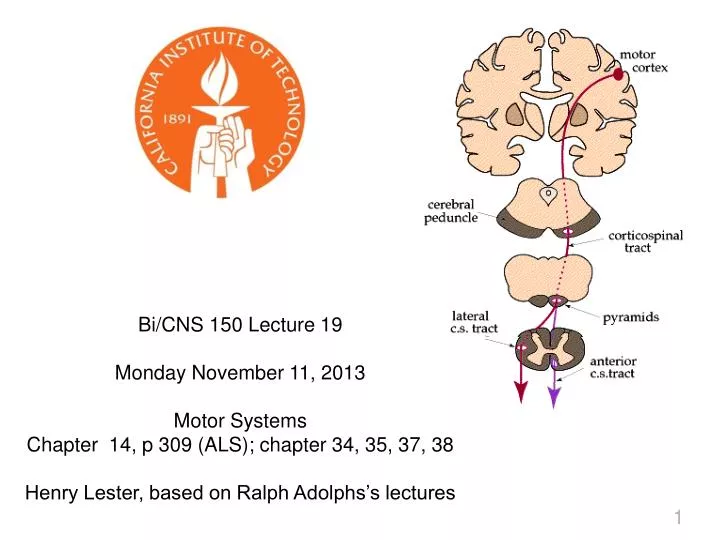
Bi/CNS 150 Lecture 19 Monday November 11, 2013 Motor Systems Chapter 14, p 309 (ALS); chapter 34, 35, 37, 38 Henry Lester, based on Ralph Adolphs’s lectures. Today: Motor cortex Corticospinal tract Motor neurons Reflexes Basal ganglia Higher motor functions. Stages of Processing.

E N D
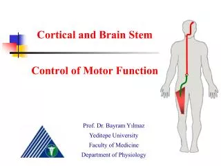
Bi/CNS 150 Lecture 19 Monday November 11, 2013 Motor Systems Chapter 14, p 309 (ALS); chapter 34, 35, 37, 38 Henry Lester, based on Ralph Adolphs’s lectures
Today: Motor cortex Corticospinal tract Motor neurons Reflexes Basal ganglia Higher motor functions
Stages of Processing 1. Transduction 2. Perception (early) 3. Recognition (late perception) 4. Memory (association) 5. Judgment (valuation, preference) 6. Planning (goal formation) 7. Action
Sensory & Motor Aspects of Behavior Account for Roughly Equal Times 4
Examples of motor output • Spinal reflexes and motor units • Posture and muscle tone • Locomotion • Control of distal extremities • Breathing • Eye movements • Speech • Emotions • Autonomic Nervous System (visceromotor) 5
Motor output at different levels Reflexes --spinal --central "Fixed action patterns" Emotional reactions Actions Long-term plans Stimulus-coupled Stimulus-decoupled 6
Motor Areas of Cortex Primary Motor Cortex BA 4 Premotor/supplementary Motor cortex BA 6 Frontal Eye Fields BA 8 Prefrontal Cortex (Frontal Association Areas) Broca’s Area (left side) BA 44, 45
Structure of Motor Cortex vs Sensory Cortex have striking differences 8
Motor System Hierarchy Motor System Hierarchy ganglia 9
Key Motor Tracts Decussation in hindbrain 10
Some Spinal Cord Motor Concepts • Motor unit: motoneuron and all innervated muscle fibers; variable number of fibers, depending on force required • Alpha-motoneuron: final common pathway • Motoneuron terminals, endplates, muscle action potentials, muscle contraction • When MN fires, all muscle fibers contract • Recruitment: adding muscle units to increase force of contraction 11
Motoneuron in Typical Spinal Cord Cross Section Dorsal Horn Sensory Myelin Motoneuron Ventral Root Motor 14 Ventral Horn Motor
Electrophysiology of the Motor Neuron and Muscle Fiber Previous Lectures 15
Herniated Disks Compress Nerve Roots (L5 most common) 16
Motor Unit Size & Physiology • Force increased by recruiting motor units • Motoneurons of different sizes: small MNS to small, slow motor units; large MNs to large, fast motor units • Size principle: smallest motor units (and smallest force) first; then larger motor units • Muscle fibers: slow (red); fatigue resistant (intermediate); fast, fatigue (white) 17
97% of spinal cord neurons are interneurons. Reflexes must be coordinated; this is complex • Sensorimotor integration in absence of supraspinal input • Motoneurons get input from sensory fibers, interneurons and descending fibers • Stretch reflexes • Flexion-withdrawal reflex • Crossed extensor reflex Groups of interneurons Tracts 19
Ipsilateral part of the crossed extensor reflex: Interneurons inhibit extensors when the flexors are commanded, and vice-versa Figure 35-2B 20
1. Sensory Organs in Muscle Participate in the Feedback Loop Intrafusal fibers in parallel with extrafusal muscle fibers Two types of sensory fibers – primary (Group Ia fibers) and secondary (Group II fibers) spindle afferents Group Ia – change in length (dynamic) Group II – length (static) Golgi tendon organ measures tension of muscle contraction Sensory information goes to spinal cord segment, dorsal column nuclei (proprioception), and cerebellum Extrafusal fibers 22
2. Gamma motoneurons in muscle participate in the feedback loop Small MNs that project out ventral roots to intrafusal fibers Activity in gamma-MNs contracts the intrafusal muscles and makes the spindle apparatus more sensitive In turn, the group Ia and II fibers become more active Gamma-bias impacts muscle tone Extrafusal fibers 23
Damage to Motoneuron (Cell body or axon) Example: Amyotrophic lateral sclerosis (ALS) “Lou Gehrig’s Disease” “Upper” motoneurons also degenerate Loss of motor unit innervation leads to weakness or paralysis of muscle Fasciculations (spontaneous contractions of muscle fibers); detected with electromyography (EMG) Atrophy of muscles, due to loss of trophic factors from motoneuron Hyporeflexia or areflexia Average time from diagnosis to death ~ 3 yr 24
The Basal Ganglia and ventral midbrain: Most Nuclei are GABAergic “striatum” Glutamatergic Dopaminergic. Future lecture on Parkinson’s disease 25
The Basal Ganglia: Major inputs “striatum” 26
Behaviors in Basal Ganglia Diseases • Three common characteristics: • tremor and other involuntary movements • changes in posture and muscle tone • slowness of movement without paralysis • Cause either excess or diminished movement • Cognitive changes (via caudate nucleus) 28
Stimulation in human motor cortex. An array is implanted . . . to localize an epileptic focus 30
Anterior Cingulate Cortex Lesions in this region cause impairment in one of the hierarchically highest levels of the motor system: the will to act . Patients with lesions to ACC can exhibit "akinetic mutism": they are not paralyzed and are conscious but respond poorly to their surroundings. They sometimes respond to very automatic things, like picking up a phone that rings next to their bedside (but then say nothing). They often recover, and then explain that while in this state, they were fully conscious but just lacked motivation to do anything and so did not respond or act on their surroundings. 31
Links Between Perception and Action: Why Can’t You Tickle Yourself?
Mirror Neurons Links Between Perception and Action: Mirror Neurons 33