Download
1 / 39
410 likes | 1.32k Views
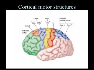
Cortical and Brain Stem Control of Motor Function. Prof. Dr. Bayram Yılmaz Yeditepe University Faculty of Medicine Department of Physiology. Motor Cortex and Corticospinal Tract. Primary motor cortex Premotor area Supplementary motor area. Motor Cortex. Premotor Area.

E N D
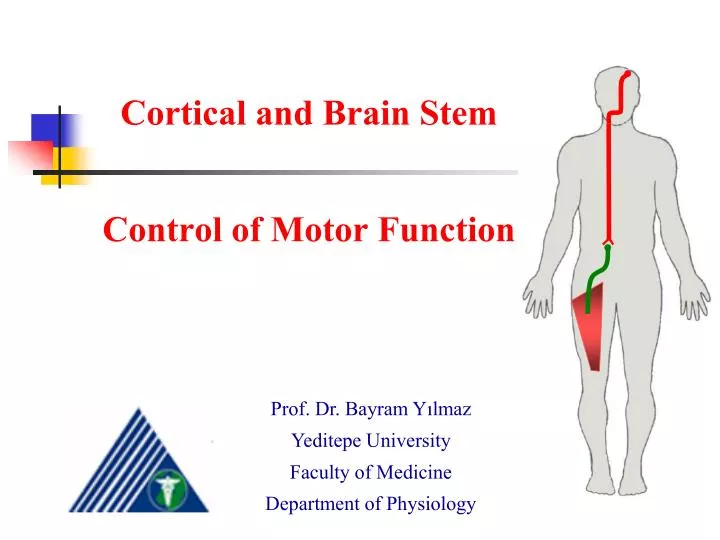
Cortical and Brain Stem Control of Motor Function Prof. Dr. Bayram Yılmaz Yeditepe University Faculty of Medicine Department of Physiology
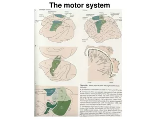
Motor Cortex and Corticospinal Tract • Primary motor cortex • Premotor area • Supplementary motor area
Premotor Area • Responsible for more complex patterns of movement • Posterior part of the premotor area sends its signals to either directly to the primary motor cortex or by way of the basal ganglia and thalamus and then back to the PMC
Supplementary Motor Area • This area functions in concert with the PMC to provide body-wide attitudinal movements and fixation movements of the different parts of the body • Stimulation of this area casuses bilatarel movements rather than unilateral
Some Specialized Areas of Motor Cortex • Broca’s area and speech • Voluntary eye movement field • Head rotation area • Area for hand skills • Motor apraxia: uncoordinated hand movements
Transmission of signals from the motor cortex to the muscles • Corticospinal tract • Or indirectly through the accessory pathways (basal ganglia, cerebellum and various brain stem nuclei) • Corticosipnal (pyramidal) tract: • It originates about 30% from the PMC, 30% from premotor area and the supplementary motor area and 40% from the somatosensory cortex • Passing through the internal capsule, forming the pyramids of the medulla
Corticospinal (pyramidal) tract • Lateral corticospinal tracts of the cord: majority of the fibers cross in the lower medulla and terminate on the interneurons in the cord gray matter • A few fibers synapse on sensory relay neurons in the dorsal horn and a few directly on the motor neurons • Ventral corticospinal tracts pass ipsilaterally down the cord, crossing in the neck or upper thoracic region • These fibers may be concerned with control of bilateral postural movements by the supplementary motor cortex
Corticospinal (pyramidal) tract • Betz cells (giant pyramidal cells) are about 60 mm in diameter • There about 34000 Betz cells in each corticospinal tract • Total number of fibers in each corticospinal tratc is more than 1 million • So Betz cells constitute about 3% of the total http://pathology.mc.duke.edu/neuropath/nawr/motor-systems.html
Other fiber pathways from the motor cortex • Axons from Betz cells send short axons to cortex itself • Large number of fibers to caudate nucleus and putamen • A moderate number of fibers to the red nuclei of the midbrain (from there rubrospinal tract) • Fibers to reticular and vestibular nuclei (reticulospinal and vestibulospinal tracts) • Many fibers to pontile nuclei, pontocerebellar fibers to the cerebellar hemispheres • Collaterals terminate in the inferior olivary nuclei – olivocerebellar pathways to cerebellum
Incoming fiber pathways to the motor cortex • Subcortical fibers from other cortical areas • Subcortical fibers from the opposite cerebral hemisphere • Somatosensory fibers directly from the ventrobasal complex of the thalamus • Tracts from the ventrolateral and ventroanterior nuclei of the thalamus (link with cerebellum and basal ganglia) • Fibers from the intralaminar nuclei of the thalamus: these fibers control the general excitability of the motor cortex
Red nucleus: and alternative pathway • Red nucleus is located in the mesencephalon • Corticorubral tract • Rubrospinal tract: crosses in the lower brain stem and courses in the lateral columns of the spinal cord • Termination on interneurons and some on motor neurons • Function of corticoruprospinal system: • Fine control of hand and finger motor movements • Corticospinal system and rubrospinal tracts are called “lateral motor system of the cord” • Vestibuloreticulospinal system (courses medially in the cord) is called “medial motor system of the cord”
Extrapyramidal system • All portions of the brain and brainstem that contribute to the motor control but are not a part of the direct corticospinal-pyramidal system • These include pathways through • Basal ganglia • Reticular formation of the brain stem • Vestibular nuclei • And often the red nuclei
Stimulus UMN LMN Voluntary Knee Extension: Neuroanatomical Description The cell body of the upper motor neuron is located in the precentral gyrus (somatotopically organized). The axon descends through the internal capsule, decussates in the medulla, descends through the lateral column of the spinal cord and terminates in the ventral horn. The cell body of the lower motor neuron is located in the ventral horn. The axon exits the CNS via ventral rootlets of spinal nerves and innervates skeletal muscle via a peripheral nerve. Skeletal muscles contract to produce the force to extend the knee.
Vertical Columnar Arrangement of Neurons in the Motor Cortex • Each column of cells functions as a single unit, usually stimulating a group of synergistic muscles • Sometimes stimulates a single muscle • Pyramidal cells that give rise to corticospinal pathway lie in the 5th layer of the cortex • Input signals enter by way of layer 2 and 4 • Layer 6 projects to other regions of the cerebral cortex • Each column can function as an amplifier • 50-100 pyramidal cells to excite a skeletal muscle
Control of Motor Functions • Dynamic and static signals transmitted by the pyramidal neurons • Somatosensory feedback to the motor cortex helps control the precision of muscle contraction
Stimulation of the spinal motor neurons • Reciprocal innervation (inhibition) to antagonistic muscle • Cord reflexes (withdrawal, walking, postural etc) can all be activated by command signals from the motor cortex • Effect of lesions in the motor cortex or in the corticospinal pathway (stroke) • Removal of the primary motor cortex • Muscle spasticity caused by lesions that damage large areas adjacent to the motor cortex • Damage to accessory pathways from nonpyramidal portions of the motor cortex
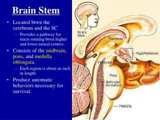
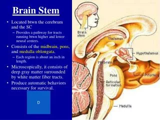
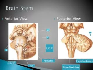
Brain Stem in Controlling Motor Function • Medulla, pons and mesencephalon • Control of respiration • Control of cardiovascular system • Partial control of GI functions • Control of many stereotyped movements of the body • Control of equlibrium • Control of eye movements • Brain stem’s reticular and vestibular nuclei
Support of the body against gravity • Pontine reticular nuclei: located posteriorly and laterally in the pons and extending into the mesencephalon • Medullary reticular nuclei
Pontine reticular system • The pontine reticular nuclei send excitatory signals downward into the cord thorugh the pontine reticulospinal tract into the anterior column of the cord • These excite axial muscles that support the body against gravity • The pontine reticular nuclei have a high degree of excitability • In addition they receive strong excitatory signals from vestibular nuclei and deep nuclei of the cerebellum
Medullary reticular system • This system sends inhibitory signals to the same anti-gravity muscles through “medullary reticulospinal tract” • This system receives strong input collaterals from • Corticospinal tract • Rubrospinal tract • Other motor pathways
Role of the vestibular nuclei to excite the antigravity muscles • The vestibular nuclei function in association with the pontine reticular nuclei to control antigravity muscles • The decerebrate animal develops spastic rigidity • Lack of input from the medullary inhibitor system
Vestibular Sensations and Equlibrium • Bony labyrinth in the temporal bone • Membranous labyrinth: functional part of the vestibular apparatus • Semicircular canals and utricle and saccule are all integral parts of the equlibrium mechanism • Macula: sensory organs of the Utricle and Saccule • Utricle: horizontal plane • Saccule: Vertical plane
Vestibular Sensations and Equlibrium • Macula is covered by a gelatinous layer • CaCO3 crystals and statoconia • There are thousands of hair cells in the macula • Bases and sides of hair cells synapse with vestibular nerve • Weight of the statoconia bends the cilia in the direction of gravitational pull • Each hair cells has 50 to 70 small cilia (stereocilia) • Kinocilium
Depolarization of Hair Cells • When stereocilia bend in the direction of kinocilium, positive ions pour into the cell from the surrounding endolymphatic fluid, causing depolarization • Under resting conditions, the nerve fibers send about 100 signals / second • When the stereocilia are bent, the impulse traffic increases • In each macula, each of the hair cells is oriented in a different direction…
Semicircular Ducts • Anterior, posterior and horizontal (lateral) ducts • They are arranged at right angles to one another • Each semicircular duct has an enlargement (ampulla) filled with fluid called endolymph • Cupula, hair cells and ampullary crest • Hair cells, kinocilia and depolarization • Bending in the opposite direction causes hyperpolarization
Semicircular Ducts, Cupula, Hair Cells and Rotation http://www.sumanasinc.com/webcontent/animations/content/vestibular.html
Function of the Utricle and Saccule • Maintenance of static equlibrium • Stimulation of hair cells and appraisal by the brain about the position of the head • Stimulation of vestibular, reticular and cerebellar motor nerve systems • Detection of linear acceleration by the utricle and saccule maculae
Detection of head rotation by the semicircular ducts • Angular acceleration and stimulation of hair cells in the cristae ampullaris in the semicircular ducts • When the rotation stops exactly opposite effects occur
Predictive function of the semicircular duct system • The semicircular ducts do not detect that the body is off balance • They detect beginning and stopping of head rotation • Semicircular dusts system predicts that dysequlibrium is going to occur & making appropriate adjustments • Removal of the cerebellar flocconodular lobes prevents normal semicircular duct signals • Vestibular mechanisms stabilizing eyes • Vestibular nuclei and medial longitudinal fasciculus to the oculomotor nuclei
Other factors concerned with equlibrium • Neck proprioceptors (joint receptors) • Vestibular apparatus detecs the orientation and movement only of the head • Proprioreceptive and exteroceptive information • Weight distribution on feet • Importance of visual information in the maintenance of equlibrium • Slowly performed motions and role of visual signals
Neuronal connections of the vestibular apparatus with the CNS • Most of the vestibular fibers terminate in the vestibular nuclei in the brainstem • Some fibers directly pass to the brainstem reticular nuclei and cerebellum • Second order neurons from vestibular nuclei also project to cerebellum, vestibulospinal tracts, medial longitudinal fasciculus and other areas of the brainstem (particularly reticular formation) • Vestibulospinal and reticulaspinal tracts & activation of antigravity muscles
Neuronal connections of the vestibular apparatus with the CNS • The flocconodular lobes of the cerebellum are especially concerned with dynamic equlibrium signals • Signals transmitted upward in the brain from both vestibular nuclei and cerebellum by way of medial longitudinal fasciculus cause corrective movements of the eyes every time the head rotates • Medial longitudinal fasciculus or reticular tracts • Project to the cerebral cortex in the parietal lobe located deep in the sylvian fissure (primary cortical center for equlibrium)
Neuronal connections of the vestibular apparatus with the CNS