Download
1 / 49
490 likes | 655 Views
BREAST. BY PROF. MOHAMED A. EL GHARBAWI e-mail: elgharma2@yahoo.com . . OBJECTIVES. BY THE END OF THIS TALK WE SHOULD KNOW EMBRYOLOGY & ANATOMY CONGENITAL ANOMALIES

E N D
BREAST BY PROF. MOHAMED A. EL GHARBAWI e-mail: elgharma2@yahoo.com.
OBJECTIVES BY THE END OF THIS TALK WE SHOULD KNOW • EMBRYOLOGY & ANATOMY • CONGENITAL ANOMALIES • TRAUMA • ACUTE INFLAMMATIONS & ABSCESSES
EMBRYOLOGY Breast is a gland embedded in fat. It is a big lipoma in which glandular tissue is embedded. ECTODERMAL in origin 2 ectodermal ridges From Axilla to Groin ( MILK LINE) Normally disappear except at the thoracic area To form 15 -20 milk ducts and lobes (each is a collection of lobules).
MILK LINES SUPERNUMERARY NIPPLES AND ACCESSORY BREASTS MAY OCCUR ALONG THIS LINE
CONGENITAL ANOMALIES A. Anomalies of the nipple: 1. Absence of the nipple (Athelia) 2. Supernumerary nipple (Polythelia) Occurring along the MILK LINE
3. Old (Congenital) retraction of nipple • Nipple fail to develop fully • Bilateral in 25% of cases • Dating since puberty • No groove around • Complications: Difficult suckling, nipple fissuring and possible abscess • Should be differentiated from RECENT RETRACTION which may indicate cancer or inflammation • Treatment: Daily pulling of the nipple, Apply negative suction on the nipple by test tube or rubber
B.ANOMALIES OF THE BREAST 1.Amazia: Absence of breast, usually unilateral 2. Polymazia : supernumerary breast, may be at axilla, groin or thigh (milk line) May be mistaken for a lipoma if nipple is absent. 3. Micromazia: congenitally small breast, unilateral or bilateral
B.ANOMALIESOF THE BREAST 4.Diffuse Hypertrophy of the Breast: Occur at puberty May be unilateral or Bilateral May be due to abnormal sensitivity to Estrogen Hypertrophy of both stroma and fat Need reduction mammoplasty with preservation of nipple & areola
ANATOMY 1 The Gladular tissue of the breast is embedded in the subcutaneous fat (stroma) to get its rounded shape. Shape: Conical. Position: From the lateral border of the sternum to Anterior axillary line and from the level of the 2nd rib to the level of the 6th rib. Rests on: Pectoralis major muscle (Upper medial 2/3rd ) Serratus anterior muscle & External Oblique muscle (Lower lateral 1/3rd )
ANATOMY 2 Suspensory ligaments of Cooper: Fibrous bands extending from skin to the Pectoralis Fascia , it prevents fall of the breasts. These ligaments become lax with age and lactation leading to lax redundant breasts. Axillary Tail of Spence: It is an upper outer extension of the breast along the lower border of the pectoralis major to the axilla through an opening in the axillary fascia called FORAMEN OF LANGER
ANATOMY 3 Structure: A collection of acini to form a lobule. A group of lobules form a lobe. They are 15 – 20 lobes, each lobe is drained into a Lactiferous duct that open at the nipple AREOLA: It is the colored circular area surrounding the nipple.
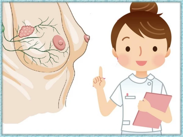
CROSS SECTION OF THE BREAST 1. CHEST WALL 2. PECTORALIS MUSCLE 3. LOBULES 4. NIPPLE 5. AREOLA 6. MILK DUCT 7. BREAST FAT 8. SKIN
ARTERIAL SUPPLY & VENOUS DRAINAGE Arterial Supply: Venous Drainage: • 1.Axillary artery Through : Superior Thoracic A. Pectoral branches of Acromio-thoracic A. Lateral Thoracic A. • 2. Internal Mammary A. • 3. 2nd,3rd and 4th intercostal Arteries (Lateral cutaneous branches). • Drain into the Axillary & Internal Mammary veins • Superficial veins anastmose with the other breast veins. • Deep veins drain to Azygos vein that communicate with the vertebral veins (AXIAL BONE METASTASES)
LYMPHATIC DRAINAGE 1: A. LYMPHATIC VESSELS: Lymph from the breast tissue is drained into 2 lymphatic plexuses: 1. Subareolar plexus of Sappy: Drains the Nipple, areola and superficial part of breast. Drained to the Deep lymphatic plexus. 2. Deep lymphatic plexus: Over the Pectoralis Fascia Drains superficial plexux, skin and deep part of breast Drained to Axillary and Internal Mammary LNs
LYMPHATIC DRAINAGE 2: B. LYMPH NODES 1: Lymph of breast drains into the Axillary & internal Mammary Lymph nodes: I. AXILLARY Lymph Nodes: They are 20-30 lymph nodes arranged in 6 groups: 1. Anterior (Pectoral)group On lateral border of pectoralis major 2. Posterior (subscapular) g. On subscapularis muscle 3. Lateral (Humeral) group on upper part of humerus 4. Intrpectoral (Rotor's) LN Between 2 pectoral Ms 5. Central (Basal) in center of axilla
LYMPHATIC DRAINAGE 3: B. LYMPH NODES 2: 6. Apical group of LNs: It is (important) as they receive lymph from the other groups. Drain directly to Jugular trunk to blood stream, so distant metastases may occur with no Supraclavicular LN affection Communicate with Supraclavicular LN Isolated Apical LNs enlarge- ment may occur as they receive lymph directly from the breast.
LYMPHATIC DRAINAGE 4: B. LYMPH NODES 3: II. INTERNAL MAMMARY LYMPH NODES: Located behind upper 4 intercostal spaces (Parasternal) Drain lymph from medial half of breast Drain into Jugular trunk and Thoracic duct Communicate with supraclavicular LNs May communicate with cutaneous lymphatics of other breast across midline. So, breast cancer may metastasize to contra lateral breast
OPERATIVE CLASSIFICATION OF LNs: THERE ARE 3 LEVELS OF AXILLARY LYMPH NODES: Level 1: Below (Lateral) to Pectoralis minor m. Level2: Behind Pectoralis minor m. Level 3: Above ( medial) to Pectoralis minor m.
OTHER LYMPHATIC CONNECTIONS: Lymphatics from inner lower quadrant communicate with sub peritoneal lymph plexus through LINEA ALBA . Malignant cells may go to peritoneum and liver. Also, through FALCIFORM LIGAMENT, malignant cells from the breast can form a nodule around the umbilicus ( SISTER JOSEPH NODULE) Malignant breast cells may migrate through the peritoneum to the pelvis and implant on the ovaries (KRUKEBERG”S TUMOUR)
HISTOLOGY:1. Breast is a glandular tissue embedded in SC fat/stroma. 2. It is formed of ducts and lobules . 3. Lobules consist of acini and form 15-20 lobes.
PHYSIOLOGY: HORMONAL CONTROL • DUCT SYSTEM: 1. Estrogen 2.Growth hormone 3. Corticosteroids • ALVEOLAR (ACINI) SYSTEM: 1.Progesterone 2. Prolactin
TRAUMATIC BREAST INJURY1 • NIPPLE CRACKING: Etiology: Trauma by teeth of baby Bad hygiene during lactation Clinical Picture: Pain &may be bleeding especially during lactation Fissure of the nipple on examination May complicate to Acute mastitis Prophylaxis: Frequent wash & dryness of nipple Use of nipple shield in presence of retracted nipple Treatment: Stop lactation from the affected breast (Avoid milk engorgement by use of suction pump) Wash & clean nipple, dry and then apply a soothing agent( Lanoline ointment ,Tincture Benzweni Co.) Apply local antibiotic cream
TRAUMATIC BREAST INJURY2 • Ecchymosis & Hematoma of breast: Due to trauma to the breast May be operative trauma Trauma may be unnoticed (-ve history) At first apply cold foments to stop and then apply hot foments to help absorption Old and calcified hematoma is hard and mimic malignancy, so excision biopsy may be indicated
TRAUMATIC BREAST INJURY3 • Traumatic Fat Necrosis: Etiology: Direct or Indirect trauma to the breast Needle biopsy or subcutaneous injection Pathology: Trauma lead to fat hydrolysis with libration Glycerol & Fatty acids. Glycerol will be absorbed. Fatty acids combine with tissue calcium forming SOAP. Soap induces foreign body reaction with aggregation of phagocytes, fibroblasts and giant cells. The formed hard mass shows Chalky white appearance of the cut surface without the yellow streaks and gritty sensation characteristic to carcinoma. Clinical picture: 50% of cases have no clear history of trauma On examination, there is a painless hard breast mass with possible nipple retraction or dimpling of the skin (suspicious of malignancy). Treatment: Excision biopsy to verify the diagnosis.
TRAUMATIC BREAST INJURY4 • Milk Fistula: Etiology: Incision (drainage of breast abscess) or injury of a milk duct in lactating breast. Rupture of a breast abscess. Clinical Picture: History of drained or neglected breast abscess. Fistula opening discharging milk. Treatment: Stop lactation. Bromocryptin (Parlodel) may be given to stop milk formation. If failed to close, fistulectomy with excision of the related sector of the gland.
TRAUMATIC BREAST INJURY5 • Traumatic Mastitis: Etiology: In females, tight brassier . In Men, tight suspender. Clinical picture: Presence of a cause. Pain and mild redness due to irritation. Treatment: Removal of the cause. • Wounds: Cut, contused or penetrating wounds according to the type of trauma. Like wounds in othe parts of the body. Repair should be cosmotic with a special attention to close dead spaces and to avoid more milk ducts.
INFLAMMATORY CONDITIOS OF BREAST • ACUTE MASTITIS: 1. Neonatal mastitis 2. Mastitis of puberty 3. Traumatic mastitis 4. Mastitis of Mumps 5. Lactation mastitis 6. Bacteria mastitis (Acute breast abscess) • CHRONIC MASTITIS: A. Non-specific 1. Chronic abscess 2. Plasma cell mastitis B. Specific 1. T.B. 2. Syphilis 3. Actinomycosis
ACUTE MASTITIS IS DISSUSSED IN THIS LECTURE CHRONIC MASTITIS WILL BE DISCUSSED IN THE NEXT LECTURE
ACUTE MASTITIS 1 • NEONATAL MASTITIS: Etiology: 1. Presence of maternal prolactin in infant blood. 2. Withdrawal of maternal estrogen from infant blood, stimulates the infant’s pituitary gland to secrete prolactin. Prolactin stimulates infant’s breast. It swells and secretes little milk). Clinical picture: 1. Swelling of the baby’s breast on 3rd or 4th day. 2. Few drops of milk is squeezed (WITCH’S MILK). 3. May be unilateral or bilateral. 4. Spontaneous subsidence on the 3rd week. Treatment: No treatment as it subside spontaneously.
ACUTE MASTITIS 2 • MASTITIS OF PUBERTY: Etiology: Disturbance of hormones at puberty. Clinical Picture: 1. May be unilateral (commoner)or bilateral. 2. Males (commoner)& Females may be affected. 3. At puberty. 4. Complaint: Pain and swelling of affected breast. 5. On Examination: Breast is swollen, tender and indurated. Never suppurate. 6. Spontaneous resolution in few weeks. Treatment: No treatment as it subsides spontaneously.
ACUTE MASTITIS 3 • TRAUMATIC MASTITIS: DISCUSSED UNDER TRAUMA TO BREAST • MASTITIS OF MUMPS: 1. A rare complication of mumps 2 Commoner in females 3. Usually unilateral 4. Spontaneous subsidence
ACUTE MASTITIS 4 • Lactation Mastitis (Milk Engorgement): Etiology: 1. During weaning from breast feeding, breasts are engorged. 2. Obstruction of lactiferous ducts by epithelial debris. 3. Non bacterial and no suppuration. Clinical Picture: 1. Occur in active lactating women. 2. Commoner in early lactation and weaning periods. 3. Complaint: Painful swollen breasts with fever due to absorption of accumulated milk protein. 4. On examination: Swollen tender breast in whole breast engorgement Or swollen and tender sector in SECTOR MASTITIS if a single lactiferous duct is obstructed.
ACUTE MASTITIS 5 • LACTATION MASTITIS (CONT.): Complication: May be infected leading to abscess. Treatment: 1. Antibiotic to protect against infection 2. Stop lactation and evacuate milk using breast pump regularly if temporary. For permanent weaning use Parlodel to stop milk formation. 3. Analgesics and antipyretics. 4. Hot foments. 5. Elevation of breast is not bacterial. 6. If suppuration happed, deal with the case as Acute bacterial mastitis ( Acute Breast Abscess).
ACUTE MASTITIS 6 • ACUTE BACTERIAL MASTITIS ( ACUTE BREAST ABSCESS)1: Etiology: A. Predisposing factors: Milk engorgement Fissure or cracks of nipple Bad hygiene of nipple & areola B. Organisms: Staph. Aureus ( Commoner/ localized inflammation). Streptcocci (Less common/ Diffuse inflammation). C .Route of Infection: Direct (Common), through fissure or crack of nipple. Lymph or Blood born (rare) D. Incidence: Common in lactating women especially at early lactation and weaning periods.
ACUTE MASTITIS 7 • ACUTE BACTERIAL MASTITIS (ACUTE BREAST ABSCESS)2: Pathology: A. Stages: Pass in 2 stages. 1. Cellulitis (pre- suppurative) stage. 2. Abscess ( suppurative) stage. B. Sites of breast abscess: 1. Retro mammary ( sub mammary). Behind the breast, on pectoral fascia. 2. Intra mammary ( in the breast tissue) 3. Pre mammary (Supra mammary) Under the skin.
ACUTE MASTITIS 8 • ACUTE BACTERIAL MASTITIS (ACUTE BREAST ABSCESS)3: Clinical Picture: Commonly in a lactating woman, especially early in lactation or in the weaning period. A. Cellutitis Stage: Symptoms & signs of acute inflammation. General: Fever, malaise and headache Local: Painful, red, hot and tender breast. Enlarged and tender axillary lymph nodes.
ACUTE MASTITIS 9 • ACUTE BACTERIAL MASTITIS (ACUTE BREAST ABSCESS)4: Clinical Picture (Cont.): B. Suppuration ( Abscess formation) Stage: Swollen, painful, red, hot and tender breast. May be subcutaneous dilated veins. Enlarged and tender axillary Lymph nodes. Pus formation is indicated by: Fever is hectic. Pain is throbbing. Edema is pitting. Fluctuation is late (don’t wait) Needle Aspiration Confirms the Diagnosis.
ACUTE MASTITIS 10 • ACUTE BACTERIAL MASTITIS (ACUTE BREAST ABSCESS)5: Complications: A. Systemic Complications: Baceraemia, septicemia, pyaemia, toxaemia. B. Damage to breast tissue. C. Chronic sinus: if neglected and ruptured under the skin. D. Chronic breast abscess ( Antibioma) is formed if antibiotics are given without drainage. Differential Diagnosis: To be differentiated from MASTITS CARCINOMATOSA ( Inflammatory Breast Cancer)
ACUTE MASTITIS 11 • ACUTE BACTERIAL MASTITIS (ACUTE BREAST ABSCESS)5: Treatment: A. In the pre supportive stage: 1. Antibiotics against Staph. & Strept ( Common). 2. Analgesic antipyretics. 3. Application of local hot foments. 4. Stop lactation with suction evacuation of milk by a breast pump. If permanent stop of lactation, give Parlodel ( Bromocryptin). 5. Breast Support ( difficult).
ACUTE MASTITIS 12 • ACUTE BACTERIAL MASTITIS (ACUTE BREAST ABSCESS)6: Treatment ( Cont. ): B. Suppurative stage: (Abscess is formed/ Must be drained/ Don’t wait for fluctuation/ Drainage should be wide enough and dependent/ under GA) Incision site: 1. Pre mammary Abscess: Incision over its pointing site. 2. Intra mammary Abscess: You may drain this abscess through a. Radial incision : from the nipple to avoid cur of lactiferous ducts. b. Circumareolar incision: Cosmetic, hidden in the border of the areola. Divide the skin only, then push a hemostat into the abscess cavity. Break the septa inside the abscess cavity by your finger to drain all pockets. c. Counter incision: To be done in the dependent position. 3. Retro mammary abscess: to be drained through the retro mammary fold .