Download
1 / 65

E N D
The development of the skull and cranial-maxillofacial deformities. Fundamentals of cranio-maxillo-facial surgery. Examination and preparation of patients for surgery. Biological basis of bone formation. Bone and cartilage grafts, implants. The principles of their application. Regeneration of the bone of the jaws. Osteogenic and osteoinductive therapy.
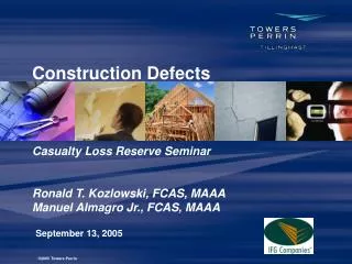
Autologus Grafts Cranium Tibia Ribs Scapula Sternum Fascia
What are craniofacial anomalies? Craniofacial anomalies (CFA) are a diverse group of deformities in the growth of the head and facial bones. Anomaly is a medical term meaning "irregularity" or "different from normal." These abnormalities are congenital (present at birth) and there are numerous variations - some are mild and some are severe and require surgery.
What causes craniofacial anomalies? • Most medical professionals agree that there is no single factor that causes these types of abnormalities. Instead, there are many factors that may contribute to their development, including the following:
combination of genesA child may receive a particular combination of gene(s) from one or both parents, or there may be a change in the genes at the time of conception, which results in a craniofacial anomaly.
environmentalThere is no data that shows a direct correlation between any specific drug or chemical exposure causing a craniofacial anomaly. However, anyprenatalexposureshouldbeevaluated.
folic acid deficiencyFolic acid is a B vitamin found in orange juice, fortified breakfast cereals, enriched grain products, and green, leafy vegetables. Studies have shown that women who do not take sufficient folic acid during pregnancy, or have a diet lacking in folic acid, may have a higher risk of having a baby with certain congenital anomalies, including cleft lip and/or cleft palate.
What are the most common types of craniofacial anomalies? • Some of the most common types of craniofacial anomalies include the following: • cleft lip and/or cleft palate - a separation that occurs in the lip or the palate (roof of the mouth), or both. Cleft lip and cleft palate are the most common congenital craniofacial anomalies seen at birth.
cleft lip - an abnormality in which the lip does not completely form. The degree of the cleft lip can vary greatly, from mild (notching of the lip) to severe (large opening from the lip up through the nose). • cleft palate - occurs when the roof of the mouth does not completely close, leaving an opening that can extend into the nasal cavity. The cleft may involve either side of the palate. It can extend from the front of the mouth (hard palate) to the throat (soft palate). Thecleftmayalsoincludethelip.
craniosynostosis - a condition in which the sutures (soft spots) in the skull of an infant close too early, causing problems with normal brain and skull growth. Premature closure of the sutures may also cause the pressure inside of the head to increase and the skull or facial bones to change from a normal, symmetrical appearance.
hemifacialmicrosomia - a condition in which the tissues on one side of the face are underdeveloped, affecting primarily the ear (aural), mouth (oral), and jaw (mandibular) areas. Sometimes, both sides of the face can be affected and may involve the skull, as well as the face. Hemifacialmicrosomia is also known as Goldenhar syndrome, brachial arch syndrome, facio-auriculo-vertebral syndrome (FAV), oculo-auriculo-vertebral spectrum (OAV), or lateral facial dysplasia.
vascular malformation - a birthmark or a growth, present at birth, which is composed of blood vessels that can cause functional or aesthetic problems. Vascular malformations may involve multiple body systems. There are several different types of malformations and they are named according to which type of blood vessel is predominantly affected. Vascular malformations are also known as lymphangiomas, arteriovenous malformations, and vascular gigantism.
hemangioma - a type of birthmark; the most common benign (non-cancerous) tumor of the skin. Hemangiomas may be present at birth (faint red mark) or appear in the first months after birth. A hemangioma is also known as a port wine stain, strawberry hemangioma, and salmon patch.
deformational (or positional) plagiocephaly - a misshapen (asymmetrical) shape of the head (cranium) from repeated pressure to the same area of the head. Plagiocephaly literally means "oblique head" (from the Greek "plagio" for oblique and "cephale" for head).
Cartilage and Bone • Cartilage--function, types, location • Bone Tissue--structure, types • Long Bone Structure and Development • Most common bone problems • Fractures • Osteoporosis
What is cartilage? • Skeletal tissue--maintains certain shape and form • Very resilient (bouncy or rubbery), mostly water • Grows fast--forms embryonic skeleton
Kinds of cartilage • Hyaline cartilage--most common, found in joints • Elastic cartilage--epiglottis, ear • Fibrocartilage--annular fibrosis of intervertebral disk, menisci of knee
Bones provide: • Support and movement (limbs, axial skeleton) • Protection (skull bones) • Mineral storage • Blood cell development (long bone marrow) Bone is made up of: 35% collagen, ground substance and cells 65% inorganic calcium (hydroxyapetite)
Bone is alive!! Bone cell types: • Osteoblasts: Make and deposit components of bone extracellular matrix • Osteoclasts: Degrade and resorb bone for remodeling • Osteocytes: “watcher cells” Sit in bone and monitor its current status
Compact Bone Dense tissue at surface of bones Haversian canals Osteocytes in lacunae Highly vascularized Fig. 6.6, p. 138 Types of bony tissue
Spongy bone Trabeculae (oriented to give mechanical strength) Interior of long bones, skull bones Epiphyses of long bones Intramembranous ossification (osteoblasts lay down bone around blood vessels in connective tissues of dermis (after 8 weeks of development) Types of bony tissue
Endochondral Ossification Fig. 6.9, p. 141 • Cartilage model • Bone collar forms in diaphysis (dense bone) • Cartilage chondrocytes in center of diaphysis die and cartilage disintegrates • Periosteal bud enters diaphysis • Makes spongy bone at ends of diaphysis (primary ossification center) • Epiphysis begins to ossify (secondary ossification center) • Hyaline cartilage remains only at • Epiphyseal surfaces (articular surfaces of joints) • Epiphyseal growth plates between diaphysis and epiphysis (primary and secondary ossification centers on either side)
“Dig holes” with hydrochloric acid Degrades calcium Phagocytize collagen fibers and dead osteocytes Line tubes (Haversian canals) left by osteoclasts Lay down new bone in circular concentric lamellae Unique to warm-blooded animals--dinosaurs??? Osteoclasts Osteoblasts
Bone Fractures • Treatment is reduction • Closed--set in place by physical manipulation from outside body • Open--surgical placement of pins or screws • Healing • Hematoma • Fibrocartilaginous callus • Bony calllus • Remodeling by osteoclasts/osteoblasts • Types of Fractures
Preoperative Preparation for Surgery • Introduction • To obtain satisfactory results in general surgery requires a careful approach to the preoperative preparation of patients . • Specific patient groups have specific needs. • High risk patients should be identified early and appropriate measures taken to reduce complications.
Overview The preoperative consultation and evaluation is an important interaction between the patient and the physician. It allows the surgeon to: carefully assess the medical condition; evaluate the patient's overall health status; determine risk factors against the procedure; educate the patient & discuss the procedure in detail.
It helps the patient to: gain a realistic understanding of the proposed surgery; consider alternative treatment options & realize the possible complications during the preoperative period. The additional time invested in a preoperative evaluation yields an improved patient physician relationship and reduces surgical complications.
Preoperative Preparation for Surgery Prior to consideration of surgical intervention, it is necessary to prepare the patient as fully as possible so as to optimize him according to his co-morbities if any The extent of pre-operative preparation will depend on the:
situation Nature of surgery (minor or major) Pre-operative preparation Facilities available Location of surgery
Preoperative Preparation for Surgery • Situation • Emergency :life-threatening condition requiring immediate action,(e.g. ruptured aneurysm, penetrating trauma) • Urgent: surgery required within a few hours (e.g. appendicitis , wound debridement) • Elective (e.g. hernia ,varicose vein)
Preoperative Preparation for Surgery • Anticipate difficulties • Make advanced preparations and organize facilities, equipment and expertise • Enhance patient safety and minimize chances of errors • Relieve any relevant fear/anxiety perceived by patient The rational for pre-operative preparation is to:
Routine Preoperative Preparation for Surgery History Physical examination Special investigation Informed consent Marking the site/side of operation Thromboembolic prophylaxis Antibiotic prophylaxis
Surgical History • History taking is detective work. • Preconceived ideas, snap judgment and hasty conclusions have no place in this process. • Do not be in any doubt that a good history is not vital. • If you embark on surgical treatment concentrating on a localized lesion you will be unprepared if complications developed. • If you take the wrong diagnostic path all the rest of your activities get misdirected.
Surgical history Presenting complaint dictates urgency, it can influence anesthetic management & any associated systemic effects of presenting pathology. Systemic assessment carefully assess each body system about its function to rule out if any other system is involved.
Surgical history • Past medical & surgical HxMany diseases have direct effect on the general and anesthetic treatment and outcome ( cardiovascular and respiratory system) Any previous operation or bleeding tendency Any previous reaction to anesthetic agent Drugs &allergic Hx Interaction with anesthesia (MAOI) Related with sudden withdrawal (steroids) Drugs for HTN ,IHD to be continued over preoperative period Anticoagulant drugs (aspirin, warfarin) HRT
Surgical history • Short-term nicotin inc. myocardial o2 demand &co dec o2 delivery • Long-term dec immune function , dec clearance of secretion Family Hx Malignant hyperthermia Pseudochlinesterase deficiency. SCA Bleeding disorders Social Hx Smoking
The physical examination Surgical signs may change and others may miss important pathology.What mind doesn't know eyes don’t see. • This includes a full physical Ex. • Don’t rely on the examination of others • One should acquire the habit of performing a complete Examination in exactly in the same sequence; • No step is omitted and added advantage of familiarizing what is normal so that abnormalities can be more recognized
The physical examination If u don’t put your finger u will put your foot General Ex. Including vitals (BP, Pulse, RR, Temp) Cardiac Ex.(JVP, HS) Respiratory Ex.(trachea, accessory ms, percussion ,auscultation) Abdominal Ex. CNS Musculoskeletal Ex Peripheral vasculature Local Ex Body orifices
The emergency physical examination Loss of life is more important than loss of a limb. The routine examination must be altered to fit the circumstances A,B,C,D,E,F Secondary survey(head-to-toe) When a number of emergencies present at the same time you must apply triage
Pre-operative investigations It is worthwhile when its requested in order to answer a specific question or confirm an important clinical impression prior to intervention Investigations are performed for a number of reasons ,but all should share the feature of directing management
Confirmation of dx Exclusion of alternative dx Pre-operative investigations To know the extent of disease Medicolegal consideration Risk to others Assessment of fitness for surgery
Pre-operative investigations: Blood tests • Full blood count (when to perform ?) • All emergency Pre-operative cases • All elective Pre-operative cases over 60 years • All elective Pre-operative cases in adult females • If surgery likely to result in significant blood loss • Suspicion of blood loss, anemia,sepsis,CRD,coagulation problems
Pre-operative investigations Blood tests Incidence of unexpected abnormality in apparently fit pt under 40 yr’s is <1% • Urea &electrolytes(when to perform?) • All Pre-operative cases over 65 • All patient with cardiopulmonary disease. Or taking diuretics, steroids • All patients with H/O renal/liver disease or abnormal nutritional state • All patients with H/O diarrhea/vomiting or other metabolic/endocrine dis. • All patients with IVF for more than 24hr’s
Pre-operative investigations Blood tests • Amylase • Perform in all adult emergency admissions with abdominal pain, prior to consideration of surgery. • RBG • Acute abdomen • Elective cases with DM, malnutrition, obesity • Elective cases over 60
Pre-operative investigations Blood tests • Clotting ; • PT inc. • with warfarin , • liver diseases; , • DIC • APTT inc • Heparin • Hemophilia A and B • BT inc • .platelet low or dysfunction • Liver function tests • All patients with upper abdominal pain ,jaundice, hepatic disease • Alcoholic • Screening for Hepatitis B and Hepatitis C
Pre-operative investigations Blood tests • Group and save /cross match • Emergency pre-operative case • Suspicion of blood loss, anemia, coagulation defects • Procedure on pregnant ladies