Download
1 / 26
260 likes | 266 Views
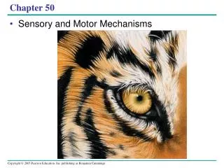
Chapter 49: Sensory and Motor Mechanism. Presented by: McQuade and Verpooten. Muscles move skeletal parts by contracting The action of a muscle Is always to contract. Human. Grasshopper. Extensor muscle relaxes. Biceps contracts. Tibia flexes. Flexor muscle contracts. Triceps relaxes.

E N D
Chapter 49: Sensory and Motor Mechanism Presented by: McQuade and Verpooten
Muscles move skeletal parts by contracting • The action of a muscle • Is always to contract
Human Grasshopper Extensormusclerelaxes Bicepscontracts Tibiaflexes Flexormusclecontracts Tricepsrelaxes Forearmflexes Extensormusclecontracts Tibiaextends Bicepsrelaxes Forearmextends Flexormusclerelaxes Triceps contracts • Skeletal muscles are attached to the skeleton in antagonistic pairs • With each member of the pair working against each other Figure 49.27
Muscle Bundle ofmuscle fibers Nuclei Single muscle fiber (cell) Plasma membrane Myofibril Z line Lightband Dark band Sarcomere TEM 0.5 m A band I band I band M line Thickfilaments(myosin) Thinfilaments(actin) H zone Z line Z line Sarcomere Vertebrate Skeletal Muscle • Vertebrate skeletal muscle • Is characterized by a hierarchy of smaller and smaller units Figure 49.28
A skeletal muscle consists of a bundle of long fibers • Running parallel to the length of the muscle • A muscle fiber • Is itself a bundle of smaller myofibrils arranged longitudinally
The myofibrils are composed to two kinds of myofilaments • Thin filaments, consisting of two strands of actin and one strand of regulatory protein • Thick filaments, staggered arrays of myosin molecules • Skeletal muscle is also called striated muscle • Because the regular arrangement of the myofilaments creates a pattern of light and dark bands
Each repeating unit is a sarcomere • Bordered by Z lines • The areas that contain the myofilments • Are the I band, A band, and H zone
The Sliding-Filament Model of Muscle Contraction • According to the sliding-filament model of muscle contraction • The filaments slide past each other longitudinally, producing more overlap between the thin and thick filaments
0.5 m (a) Relaxed muscle fiber. In a relaxed muscle fiber, the I bandsand H zone are relatively wide. Z H A Sarcomere (b) Contracting muscle fiber. During contraction, the thick andthin filaments slide past each other, reducing the width of theI bands and H zone and shortening the sarcomere. (c) Fully contracted muscle fiber. In a fully contracted musclefiber, the sarcomere is shorter still. The thin filaments overlap,eliminating the H zone. The I bands disappear as the ends ofthe thick filaments contact the Z lines. • As a result of this sliding • The I band and the H zone shrink Figure 49.29a–c
The sliding of filaments is based on • The interaction between the actin and myosin molecules of the thick and thin filaments • The “head” of a myosin molecule binds to an actin filament • Forming a cross-bridge and pulling the thin filament toward the center of the sarcomere
Thick filament Thin filaments 1 Starting here, the myosin head is bound to ATP and is in its low-energy confinguration. 5 Binding of a new mole- cule of ATP releases the myosin head from actin, and a new cycle begins. Thin filament Myosin head (low-energy configuration) The myosin head hydrolyzes ATP to ADP and inorganic phosphate ( I ) and is in its high-energy configuration. ATP 2 ATP Cross-bridge binding site Thick filament P Actin Thin filament moves toward center of sarcomere. Myosin head (high-energy configuration) ADP Myosin head (low-energy configuration) P i 1 The myosin head binds toactin, forming a cross-bridge. 3 ADP + Cross-bridge ADP P i P i Releasing ADP and ( i), myosinrelaxes to its low-energy configuration, sliding the thin filament. 4 P • Myosin-actin interactions underlying muscle fiber contraction Figure 49.30
Tropomyosin Ca2+-binding sites Actin Troponin complex (a) Myosin-binding sites blocked • When a muscle is at rest • The myosin-binding sites on the thin filament are blocked by the regulatory protein tropomyosin Figure 49.31a
Ca2+ Myosin-binding site (b) Myosin-binding sites exposed • For a muscle fiber to contract • The myosin-binding sites must be uncovered • This occurs when calcium ions (Ca2+) • Bind to another set of regulatory proteins, the troponin complex Figure 49.31b
Motorneuron axon Mitochondrion Synapticterminal T tubule Sarcoplasmicreticulum Ca2+ releasedfrom sarcoplasmicreticulum Myofibril Sarcomere Plasma membraneof muscle fiber • The stimulus leading to the contraction of a skeletal muscle fiber • Is an action potential in a motor neuron that makes a synapse with the muscle fiber Figure 49.32
The synaptic terminal of the motor neuron • Releases the neurotransmitter acetylcholine, depolarizing the muscle and causing it to produce an action potential
Action potentials travel to the interior of the muscle fiber • Along infoldings of the plasma membrane called transverse (T) tubules • The action potential along the T tubules • Causes the sarcoplasmic reticulum to release Ca2+ • The Ca2+ binds to the troponin-tropomyosin complex on the thin filaments • Exposing the myosin-binding sites and allowing the cross-bridge cycle to proceed
Acetylcholine (ACh) released by synaptic terminal diffuses across synapticcleft and binds to receptor proteins on muscle fiber’s plasma membrane, triggering an action potential in muscle fiber. Synapticterminalof motorneuron 1 PLASMA MEMBRANE Synaptic cleft T TUBULE Action potential is propa- gated along plasma membrane and down T tubules. 2 ACh SR 4 Action potential triggers Ca2+ release from sarco- plasmic reticulum (SR). 3 Ca2 Calcium ions bind to troponin; troponin changes shape, removing blocking action of tropomyosin; myosin-binding sites exposed. Tropomyosin blockage of myosin- binding sites is restored; contraction ends, and muscle fiber relaxes. 7 Ca2 CYTOSOL Cytosolic Ca2+ is removed by active transport into SR after action potential ends. 6 ADP P2 Myosin cross-bridges alternately attach to actin and detach, pulling actin filaments toward center of sarcomere; ATP powers sliding of filaments. 5 • Review of contraction in a skeletal muscle fiber Figure 49.33
Neural Control of Muscle Tension • Contraction of a whole muscle is graded • Which means that we can voluntarily alter the extent and strength of its contraction • There are two basic mechanisms by which the nervous system produces graded contractions of whole muscles • By varying the number of fibers that contract • By varying the rate at which muscle fibers are stimulated
Motorunit 1 Motorunit 2 Spinal cord Synaptic terminals Nerve Motor neuroncell body Motor neuronaxon Muscle Muscle fibers Tendon • In a vertebrate skeletal muscle • Each branched muscle fiber is innervated by only one motor neuron • Each motor neuron • May synapse with multiple muscle fibers Figure 49.34
A motor unit • Consists of a single motor neuron and all the muscle fibers it controls • Recruitment of multiple motor neurons • Results in stronger contractions
Tetanus Tension Summation of two twitches Singletwitch Time Actionpotential Pair ofactionpotentials Series of action potentials at high frequency • A twitch • Results from a single action potential in a motor neuron • More rapidly delivered action potentials • Produce a graded contraction by summation Figure 49.35
Tetanus is a state of smooth and sustained contraction • Produced when motor neurons deliver a volley of action potentials
Types of Muscle Fibers • Skeletal muscle fibers are classified as slow oxidative, fast oxidative, and fast glycolytic • Based on their contraction speed and major pathway for producing ATP
Other Types of Muscle • Cardiac muscle, found only in the heart • Consists of striated cells that are electrically connected by intercalated discs • Can generate action potentials without neural input
In smooth muscle, found mainly in the walls of hollow organs • The contractions are relatively slow and may be initiated by the muscles themselves • In addition, contractions may be caused by • Stimulation from neurons in the autonomic nervous system