Download
1 / 48
490 likes | 642 Views
In the name of GOD. Direct NMR Detection of Alkali Metal Ions Bound to G- Quadruplex DNA. J. AM. CHEM. SOC. 2008, 130, 3590-3602. Zeinab Mokhtari. 15-Feb-2011. Queen’s university, Canada, founded in 1841. Ramsey Ida and Gang Wu.

E N D
In the name of GOD Direct NMR Detection of Alkali Metal Ions Bound to G-Quadruplex DNA J. AM. CHEM. SOC. 2008, 130, 3590-3602 ZeinabMokhtari 15-Feb-2011
Queen’s university, Canada, founded in 1841 Ramsey Ida and Gang Wu
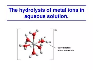
A telomere is a region of repetitive DNA at the end of a chromosome, which protects the end of the chromosome from deterioration. Its name is derived from the Greek nouns telos (τέλος) "end" and merοs (μέρος, root: μερ-) "part". The telomere regions deter the degradation of genes near the ends of chromosomes by allowing for the shortening of chromosome ends, which necessarily occurs during chromosome replication. Introduction Telomeres function by preventing chromosomes from losing base pair sequences at their ends. They also stop chromosomes from fusing to each other. Telomere When the telomere becomes too short, the chromosome reaches a "critical length" and can no longer replicate. This means that a cell becomes "old" and dies by a process called apoptosis. http://en.wikipedia.org/wiki/Telomere Telomeres do not contain the codes for proteins are not themselves genes.
Introduction J. AM. CHEM. SOC. 2003, 125, 13895-13905 (A) Atomic numbering for 5’-GMP. (B) G-quartet model. (C) Space-filling model of the G-quartet. (D) Diagram showing the two types of cation environments in 5’-GMP.
Introduction J. AM. CHEM. SOC. 2003, 125, 10830-10841
G-quadruplexes characterization solution NMR spectroscopy or crystallography Introduction
The mode of alkali metal ion binding in G-quadruplex DNA and RNA Alkali metal ions : important roles in the formation, stability, and structural polymorphism of G-quadruplex DNA and RNA Introduction high-resolution crystallography and solid-state NMR much more difficult in solution
Laszlo and coworkers 1979-1980 the first model for ion binding to a G-quartet structure solution 23Na, 39K, and 87Rb NMR Introduction
Relaxation data analysis Later, Braunlin and co-workers 1993-1996 Solution 23Na and 39K NMR techniques to directly study ion binding to G-quadruplex DNA. Introduction Conclusion : alkali metal ions tightly bound to a G-quadruplex DNA are “invisible” to NMR in solution, because of low signal intensity and unfavorable quadrupole spin relaxation properties. An indirect approach : NMR relaxation properties of alkali metal ions are measured for the averaged signal and analyzed using either two-site or three-site chemical exchange models. relaxation data analysis Only ion binding information for the tightly bound sites
Since the late 1990s NMR methodologies based on spin-1/2 directly probing NH4+and Tl+ions in G-quadruplex DNA new information about NH4+ion movementinside G-quadruplex channels Introduction
multinuclear NMR methodology Ramsey Ida and Gang Wu 2005 Alkalimetal ions (Na+, K+, and Rb+) tightly bound to a G-quadruplex structure can be directly observed by 23Na, 39K, and 87Rb NMR even in the liquid state. Introduction directly studying alkali metal ion binding to G-quadruplex DNA d(TG4T), d(G4T3G4), and d(G4T4G4)
d(TG4T) The G-quadruplex structure formed by DNA hexamerd(TG4T) in the presence of Na+ or K+ ions • The solution structure : • Four d(TG4T) strands form a parallel-stranded G-quadruplex structure containing four stacked G-quartets. • only one 23Na NMR signal at -17 ppm Introduction • The crystal structure : • Four-stranded parallel structure similar to the solution structure. • A distinct intermolecular stacking (a pair of quadruplex structures that are stacked at the 5’ ends) • A total of seven Na+ ions located either between or within the G-quartet planes crystal packing effect • Solid-state 23Na NMR : • Na+ ions between two adjacent G-quartets (signal centered at -19 ppm) • the illusive signal from the in-plane Na+ ions because of the presence of a large signal due to phosphate-bound Na+ ions
Whether the mode of Na+ ion binding is the same in solution and solid states? Introduction
Clark et al. 2003 A crystal structure of d(TG4T)complexed to a drug molecule, daunomycin Daunomycin prefers to be stacked onto the ends of the G-quadruplex rather than to intercalate between the G-quartet layers. Na+ ions are located only between adjacent G-quartet planes. Introduction
d(G4T3G4) Neidle and co-workers 2006 The crystal structures of the K+ form of d(G4T3G4) • d(G4T3G4) forms a bimolecular intermolecular G-quadruplex with two lateral thymine loops ( either on the same G-quadruplex face, forming a head-to-headdimer, or on the opposite faces, resulting in a head-to-taildimer). Introduction • K+ ions were found between G-quartets, each being equidistant from eight O6 guanine atoms. • Not large energy differences between the head-to-head and head-to-tail bimolecular G-quadruplexes containing the T3 loops • Both of these forms may be present in solution.
d(G4T4G4) The bimolecular G-quadruplex structure formed by d(G4T4G4) : related to the repeat sequence d(T4G4) found in the OxytrichanoVatelomere Solution : an antiparallel, bimolecular quadruplex structure consisting of four stacked G-quartets and two diagonal thymine loops. Introduction • Feigon and co-workers 1999 , 2003 • various ionic forms of d(G4T4G4) in solution • The nature of monovalentcations present in the solution (Na+, K+ and NH4+) does not affect the overall fold of d(G4T4G4). • In the presence of Ca2+, d(G4T4G4) can undergo a structural transition from an antiparallel to a parallel quadruplex, which in turn can aggregate into a large molecular assembly known as the G-wire.
d(G4T4G4) K+ The crystal structure of the K+form of d(G4T4G4) Very similar to that found in solution Introduction Explicit information about the mode of ion binding in d(G4T4G4) • Three K+ ions resided inside the quadruplexchannel, each being sandwiched between two adjacent G-quartets. • Two additional K+ ions in the thymine loop regions
d(G4T4G4) Tl+ the same structure as the K+ form Interestingly, when an acridine derivative interacts with d(G4T4G4), the drug molecule enters into one of the thymine loop regions, replacing the loop K+ ion in the same loop but leaving the other loop K+ ion unperturbed. Introduction • solution 205Tl NMR : • four signals between 80 and 150 ppm • Two of the four 205Tl NMR signals : Tl+ ions inside the quadruplexchannel in the same coordination environment as those seen for K+, NH4+, and Na+ • The other two signals : Tl+ ions residing in the thymine loop region
d(G4T4G4) NH4+ Three NH4+ ions are located inside the G-quadruplex channel in a way identical to the mode of K+ binding observed in the crystal structure. Introduction However, no NMR evidence was found for NH4+ ions to be located in the thymine loop region.
d(G4T4G4)-protein Na+ Crystal structure : a completely different mode of ion binding Two Na+ ions are located nearly coplanar with the two central G-quartets, and two additional Na+ ions are located in the thymine loop regions. Introduction Although these crystallographic studies have provided unequivocal evidence about the mode of K+, Tl+, and Na+ ion binding both inside the quadruplexchannel and in the thymine loop region, it is far less certain whether the same type of ion binding should occur in solution for the Na+ form of d(G4T4G4).
d(G4T4G4) Na+ solid-state 23Na NMR : three Na+ ions reside inside the G-quadruplexchannel, each being sandwiched between two adjacent G-quartets Introduction Clearly different
The exact mode of Na+ ion binding in d(G4T4G4)??? Introduction
d(TG4T), d(G4T3G4), and d(G4T4G4) In this study : Why? • The G-quadruplex structures formed from these DNA oligomers have been fully characterized by either solution NMR or crystallography. Introduction • They represent three classic types of G-quadruplexes: parallel-stranded, bimolecular with lateral loops, and bimolecular with diagonal loops. • No information is available regarding the mode of Na+ ion binding to these G-quadruplexes in solution.
Sample Preparation • DNAoligonucleotides of d(TG4T), d(G4T3G4), and d(G4T4G4) • Synthesized • Purified • For Na+ forms of DNA : Dissolved in deionized water followed by extensive dialysis against NaCl • Lyophilized • For NMR experiments : DNA oligomers were dissolved in sodium phosphate buffer with appropriate amounts of NaCl added. • strand concentrations of the DNA : UV-vis spectrometer • The total Na+ ion concentration : NaCl standard and 23Na NMR Experimental Details
NMR Experiments • NMR spectrometers • Bruker Avance-400 (9.4 T) • Avance-500 (11.7 T) • Avance-600 (14.1 T) WATERGATE sequence to suppress the water Signal in 1H NMR experiments. The pulse sequence of longitudinal eddy current delay (LED) with bipolar-gradient pulses for H DOSY experiments. Experimental Details A 5-mm quartz NMR tube for solution 23Na NMR experiments, to reduce the 23Na background signal from regular glass materials. The 1D 23Na NMR spectra were acquired using an inversion-recovery sequence with the interpulse delay (recovery delay) set to appropriate values depending on the actual T1value for the free 23Na signal. 1D 87Rb NMR spectra : single-pulse sequence Solid-state 23Na NMR spectra : BrukerAvance-II 900 (21.1 T) spectrometer using a 4-mm magic angle spinning (MAS) probe
Direct Observation of Na+ Ions inside the G-Quadruplex Channel The large number of imino1H NMR signals (ca. 16 peaks) suggests that either d(G4T3G4) forms an asymmetric dimeric G-quadruplex or two dimeric G-quadruplex structures coexist in solution. As mentioned earlier, the crystallographic study of the K+ form of d(G4T3G4) indeed suggests that head-to-tail antiparallel and head-to-head dimers are both possible. imino1H NMR signal confirming the formation of fully folded G-quadruplex Results and Discussion a well-defined signal at -17.7 ppm (Na+ ions residing inside the G-quadruplexchannel being coordinated to eight O6 guanine atoms) No solution 1H NMR structure has been reported in the literature. 1H and 23Na NMR spectra for the Na+ forms of d(TG4T), d(G4T3G4), and d(G4T4G4)
A general concern regarding 23Na NMR studies of DNA It is necessary to employ NMR techniques to suppress the large 23Na NMR signal arising from free Na+ ions, allowing much weaker signals due to DNA-bound Na+ ions to be effectively detected. inversion-recovery pulse sequence T1 filter Results and Discussion The 23Na NMR signal at δ= 0 ppm (denoted as the free Na+ signal) is actually the averaged signal for Na+ ions undergoing fast exchange between a truly free state and a phosphate-bound state. high-quality 23Na NMR spectra for DNA Because the free Na+ ions have a much longer spin-lattice relaxation time (typically T1 ~10 ms) than do the tightly bound Na+ ions (typically T1 < 1 ms), we can set the interpulse delay (recovery delay) to be very close to the so-called null (zero-crossing) point for the free Na+ ions so that the signal from the free Na+ ions would be greatly reduced, whereas the signals from the bound Na+ ions have already fully recovered at this point and thus show their full intensities in the spectra.
Na+ ion occupancy inside the G-quadruplex channel The concentration of Na+ ions that give rise to the 23Na NMR signal at -17.7 ppm in each of the three DNA samples d(G4T3G4) d(G4T4G4) three channel sites Results and Discussion straightforward to estimate the ion occupancy approximately 100% each G-quadruplex channel contains three Na+ ions two stacking tetramolecular G-quadruplexes related by symmetry not possible to assess with 1H NMR d(TG4T) 1H DOSY complicated
1H DOSY molecular translational coefficient (D) of the d(TG4T) G-quadruplex combined bead/cylinder model Not a dimer in the DNA concentrations used in our study Results and Discussion the length of the d(TG4T) G-quadruplex in D2O: 20 ± 4 Å half of the length for the G-quadruplexdimer found in the crystal structures Similar results : 3 ions per G-quadruplex
Competitive Binding of Na+ and Rb+ Ions for the G-Quadruplex Channel Site well-defined NMR signals for alkali metal ions residing inside the G-quadrupelx channel possible to study competitive ion binding by simultaneously detecting the two competing metal ions d(TG4T) Results and Discussion Rb+/Na+ titration experiment Rb+ ions undergoing fast exchange between free and phosphate-bound states First time 0 ppm 70 ppm Two 87Rb NMR signals gradually replacement of the Na+ ions Rb+ ions residing inside the G-quadruplex channel
Results and Discussion d(TG4T) Illustration of the competitive Rb+/Na+ ion binding for the channel site of d(TG4T) G-quadruplex
Confirmed by 1H NMR data Assuming full ion occupancy inside the channel and no DNA unfolding during the Rb+/Na+ titration experiment: Results and Discussion Rb+ ions residing inside the channel: fit the curve of [Rb+]channel versus [Rb+]total K
K=1.6±0.2 at 298 K for the Rb+/Na+ competition for the G-quadruplex channel site Results and Discussion Rb+ ions are preferred over Na+. This value of K is also in agreement with that reported by Wong and Wu for 5’-GMP, K=1.8 at 298 K, on the basis of a solid-state NMR titration experiment. J. AM. CHEM. SOC. 2003, 125, 13895-13905 a valid approach for obtaining solution properties The Na+ ion at the channel site is fully dehydrated no difference between measurements in solution or solid state
5’-GMP • two separate 23Na NMR signals : • -17.0 ppm : Na+ ions inside a G-quadruplexfilled with Na+ions • -16.2 ppm : Na+ ions in a channel containing mixed Rb+/Na+ions Results and Discussion channel Na+ ion signal for d(TG4T) is much broader Not observation of resolved 23Na NMR signals for the channel Na+ ions The situation is different in the K+/Na+ titration experiment.
Residence time of Na+ ions inside the channel The frequency separation between the two 87Rb NMR signals (9.2 kHz at 9.4 T) also allows us to conclude that the residence time of Rb+ ions inside the G-quadruplex channel must be much longer than 17 is at 298 K. Halle and co-workers demonstrated a magnetic relaxation dispersion (MRD) NMR approach to study competitive Rb+ and Na+binding to the minor groove of a B-DNAduplex, [d(CGCGAATTCGCG)]2 Results and Discussion Weak ion binding mean residence time for Rb+= 0.2 μs for Na+ =10 ns to 100 μs Much shorter than those estimated for the Na+ and Rb+ ions residing inside a G-quadruplex channel Averaged NMR signal was in 23Na and 87Rb NMR spectra of the B-DNA duplex Direct NMR + MRD
Observation of Na+ Ions in the T4 Loop Region of d(G4T4G4) Results and Discussion • Four regions for ion binding in G-quadruplex structure: • Phosphate • Groove • Loop • Channel Na+ ions residing in the diagonal T4 loop regions (at -7.4 ppm)
Groove Regions groove width : closest distance between two phosphate oxygen atoms across the groove Results and Discussion
Groove Regions groove width : closest distance between two phosphate oxygen atoms across the groove parallel-stranded All glycosidic bonds of the guanine bases are in anti conformation, resulting in the formation of four equally wide groove regions. 8.30 and 9.53 Å Cross-phosphate ion binding • syn-syn-anti-anti pattern in glycosidic torsion angles within each G-quartet + diagonal topology three groove regions • One narrow groove (6.76 Å) • Two medium grooves (10.59 and 10.26Å) • One wide groove (14.74 Å) Na+ ions residing in the diagonal T4 loop regions
Syn and anti conformations of guanine Biol. Chem., Vol. 382, pp. 621 – 628, April 2001
Addition of Mg2+ions to the Na+ form of d(G4T4G4) more strongly interaction with the DNA phosphates but no entering in the G-quadrupelxchannel Decreases of the line width of the 23Na signal at 0 ppm, free and phosphate-bound states, from 190 Hz before the titration experiment to 46 Hz when the concentration of Mg2+ ions reaches 4.7 mM. Results and Discussion drastic line-width reduction Mg2+ ions compete very effectively only for the DNA phosphate backbone. K+ ions are capable of competing for all three sites. -7.4 and -17.7 ppm unchanged
Na+ Ions in the T4 Loop Are Less Tightly Bound Than the Channel Ions. The loop Na+ ions are less tightly bound (or more mobile) than the channel Na+ ions, and thus they undergo a much faster exchange between the bound and free states. Results and Discussion For the channel Na+ ions, the fact that the 23Na NMR line width shows very little temperature dependence suggests a much longer residence lifetime. Figure 11. Variable-temperature 23Na NMR spectra of d(G4T4G4) (5.4 mM strand concentration and [Na+]total =70 mM).
K+ Ions Compete for Both Loop and Channel Sites of d(G4T4G4). K+/Na+ ion titration When K+ ions are added to the DNA solution, they would replace the Na+ ions already bound to the quadruplex channel sites. Results and Discussion The loop binding site also exhibits a higher affinity for K+ ions than for Na+ ions. 23Na NMR spectra of d(G4T4G4) from a K+/Na+ ion titration experiment
Direct 23Na NMR Evidence for a G-Quadruplex Channel Filled with Mixed Na+ and K+ Ions Type III : channel Na+ ion that resides at the inner site with two K+ ions occupying the two outer sites; with low concentration because of [K+]<<[Na+] Type II channel Na+ ion : the Na+ ion residing inside the quadruplex channel but having a K+ ion at the nearest binding site Results and Discussion
previous studies of G-quadruplex channel with mixed ions X-ray crystallographic or solution 1H and 15NH4+ NMR data Caceres et al. : two crystal structures for d(TG4T) with mixed Tl+ and Na+ ions Neidle and coworkers : mixed Ca2+ and Na+ ions in the d(TG4T) quadruplex Results and Discussion Sundaralingam and co-workers : mixed Ba2+ and Na+ ions in a RNA quadruplex formed by (BrdU)r- (GAGGU). Plavec and co-workers : 1H NMR evidence for the presence of mixed NH4+ and K+ (or Na+) ions in d(G3T4G4). • Mixed ion information • Remarkable sensitivity of the 23Na NMR chemical shift to subtle difference in ion coordination environment solution 23Na NMR
A Hypothesis on MonovalentCation Binding in Diagonal T4 Loops The loop ion is located above the terminal G-quartet plane, coordinating to four O6 atoms from the terminal G-quartet, two O2 atoms from the loop thymine bases (T5 and T7), and two water molecules in a square anti-prism geometry. Results and Discussion K+_O distances considerably longer than the corresponding Na+_O values Molecular model of the Na+ & K+ ion binding site in the T4 loop of d(G4T4G4)
Conclusion Na+ and Rb+ ions residing inside G-quadruplex channel are “NMR visible” in solution. Competitive ion binding to a particular site in DNA can be directly monitored by simultaneous NMR detection of the two competing metal ions. Monovalentcation binding to the diagonal T4 loop of a G-quadruplex may be a general phenomenon. Solution multinuclear NMR of alkali metals is a viable technique for studying alkali metal ion binding to DNA.