Download
1 / 1
10 likes | 186 Views
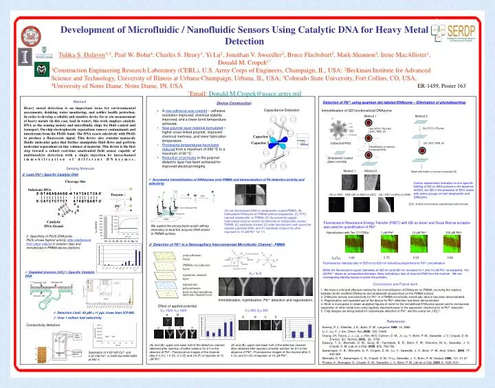
A. B. C. Capacitor 1. D. E. F. Capacitor 2. Fe 3+. V 1. V 1. PCTE membrane. ground. ground. V 2. ground. separation channel. ground. detection channel. polycarbonate block. Enzyme. Pb 2+. PMMA via reduction layer. separation channel layer. nanoporous polycarbonate

E N D
A B C Capacitor 1 D E F Capacitor 2 Fe3+ V1 V1 PCTE membrane ground ground V2 ground separation channel ground detection channel polycarbonate block Enzyme Pb2+ PMMA via reduction layer separation channel layer nanoporous polycarbonate track-etched membrane detection channel layer Development of Microfluidic / Nanofluidic Sensors Using Catalytic DNA for Heavy Metal Detection Tulika S. Dalavoy1,2, Paul W. Bohn4, Charles S. Henry3, Yi Lu2, Jonathan V. Sweedler2, Bruce Flachsbart2, Mark Shannon2, Irene MacAllister1, Donald M. Cropek1* 1Construction Engineering Research Laboratory (CERL), U.S. Army Corps of Engineers, Champaign, IL, USA; 2Beckman Institute for Advanced Science and Technology, University of Illinois at Urbana-Champaign, Urbana, IL, USA; 3Colorado State University, Fort Collins, CO, USA, 4University of Notre Dame, Notre Dame, IN, USA *Email: Donald.M.Cropek@usace.army.mil ER-1459, Poster 163 Abstract Heavy metal detection is an important issue for environmental assessments, drinking water monitoring, and soldier health protection. In order to develop a reliable and sensitive device for in situ measurement of heavy metals (in this case, lead in water), this work employs catalytic DNA as the sensing moiety and microfluidic chips for fluid control and transport. On-chip electrophoretic separations remove contaminants and interferents from the Pb(II) band.The DNA reacts selectively with Pb(II) to produce a fluorescent signal. This device also contains nanoscale fluidic molecular gates that further manipulate fluid flows and perform molecular separations on tiny volumes of material. This device is the first step toward a robust real-time unattended field sensor capable of multianalyte detection with a single injection by intrachannel immobilization of different DNAzymes. Sensing Molecule Detection of Pb2+ using quantum dot labeled DNAzyme – Elimination of photobleaching Device Construction Capacitance Detection Immobilization of QD functionalized DNAzyme • A new adhesive was created – adhesive resolution improved, chemical stability improved, and a lower bond temperature achieved. • New polymer layer material formulated – higher cross-linked polymer, improved chemical inertness, and lower fabrication temperature. • Processing temperatures have been reduced from a maximum of 200 °C to a maximum of 90 °C. • Reduction of pinholes in the polymer dielectric layer has been achieved for improved electrical integrity. Method 1 Method 2 Bio-5T(7)-17Ea-Am Bio-5T(7)-17Ea-Am EDC, PBS, 2h CdSe/ZnS/PEG QD, EDC, PBS, 2h Floating ON, 40s Ultrafiltration to remove unreacted DNA ON Streptavidin coated glass coverslip Method 1 Method 2 Wash with buffer to remove unreacted QD Lead (Pb2+) Specific Catalytic DNA OFF OFF, 4s OFF, 30s • Successful immobilization of DNAzymes onto PMMA and demonstration of Pb detection activity and selectivity Cleavage Site Control experiments indicates no non-specific binding of QD on SSG surface in the absence of EDC, but QD in the presence of EDC reacts with amine groups on both streptavidin and DNAzyme. Substrate DNA QD on SSG DNA +QD on SSG (no EDC) QD + EDC on SSG (no DNA) Pb2+ EDC- N-ethyl aminomethyl carbodiimide hydrochloride ON OFF Biotin-DNAzyme-FAM (A) non-biotinylated DNA on streptavidin-coated PMMA; (B) biotinylated DNAzyme on PMMA without streptavidin; (C) FITC-labeled streptavidin on PMMA; (D) fluorescently-tagged, biotinylated enzyme strand immobilized on streptavidin-coated PMMA; (E) substrate of panel (D) after hybridization with quencher-labeled substrate DNA; and (F) substrate of panel (E) after exposure to 10 mM Pb2+ for 1 h. streptavidin PMMA Catalytic DNA Strand Fluorescence Resonance Energy Transfer (FRET) with QD as donor and Texas Red as acceptor was used for quantification of Pb2+ We exploit the strong biotin-avidin affinity chemistry to bind the enzyme DNA strand to PMMA surface. Hybridization with Tex-(7)17DSa 1 μM Pb2+ 10 μM Pb2+ 100 μM Pb2+ • Specificity of Pb(II) DNAzyme. • Pb(II) shows highest activity, little interference from other cations in solution (top) and immobilized in PMMA device (bottom). Detection of Pb2+ in a Nanocapillary Interconnected Microfluidic Channel - PMMA I525/I620 0.66 0.73 0.93 0.68 V2 Fluorescence intensity ratio of 525 nm to 620 nm should be proportional to Pb2+ concentration. While the fluorescence signal intensities at 525 nm and 620 nm increase for 1 and 10 mM Pb2+ as expected, 100 mM Pb2+ shows an unexpected decrease, likely indicating a loss of enzyme DNA from the channel. We are investigating stability issues to solve this problem. • Depleted Uranium (UO22+) Specific Catalytic DNA V2 > V1/2 Conclusions and Future work 1. We have a mild and effective method for the immobilization of DNAzyme on PMMA, involving the reaction between biotin-modified DNAzyme and streptavidin physisorbed on the PMMA surface. 2. DNAzyme activity and selectivity for Pb2+ in a PMMA microfluidic-nanofluidic device has been demonstrated. 3. Regeneration and repeated use of the device for Pb2+ detection has been demonstrated. 4. Work is in progress to obtain analytical figures of merit for the immobilized DNAzyme system and to incorporate separation of other metal ions using capillary electrophoresis in the separation channel prior to Pb2+ detection. 5. Chip designs are being tested for multianalyte detection of Pb2+ and the uranyl ion, UO22+. Immobilization, hybridization, Pb2+ detection and regeneration. Effect of applied potential V1= 150V, V2= 100V V1= 50V, V2= 30V • Detection Limit: 45 pM = 11 ppt, lower than ICP-MS. A B A B C D • Over 1 million fold selectivity. References Conductivity detection Kemery, P. J.; Steehler, J. K.; Bohn, P. W. Langmuir1998, 14, 2884 Li, J.; Lu, Y. J. Am. Chem. Soc.,2000, 122, 10466 Chang, I-H; Tulock, J. J.; Liu, J.; Kim, W-S; Cannon, D. M., Jr.; Lu, Y.; Bohn, P. W.; Sweedler, J. V.; Cropek, D. M. Environ. Sci. Technol. 2005, 39, 3756 Dalavoy, T. S.; Wernette, D. M.; Gong, M.; Flachsbart, B. R.; Bohn, P. W.; Shannon, M. A.; Sweedler, J. V.; Cropek, D. M. Lab on a Chip, 2008, 8(5), 786-793 Swearingen, C. B.; Wernette, D. P.; Cropek, D. M.; Lu, Y.; Sweedler, J. V.; Bohn, P. W. Anal. Chem., 2005, 77, 442-448 Wernette, D. P.; Swearingen, C. B.; Cropek, D. M.; Yi Lu, Sweedler, J. V.; Bohn, P. W. Analyst, 2006, 131, 41-47 Piruska, A.; Branagan, S.; Cropek, D. M.; Sweedler, J. V.; Bohn, P. W. Lab on a Chip, 2008, 8, 1625-1631 C D E F Cu2+ (A) and (B); upper and lower half of the detection channel obtained after injection of buffer solution for 2 h in the absence of Pb2+, Fluorescence images of the channel after 0 h (C), 1 h (D), 2 h (E) and 3 h (F) of injection of 10 mM Pb2+. (A) and (B); upper and lower half of the detection channel after obtained after injection of buffer solution for 2 h in the absence of Pb2+; Fluorescence images of the channel after 0 h (C) and 2 h (D) of injection of 10 mM Pb2+. Separation of 4.68 mM Cu2+ and 5.32 mM Fe3+ in 5mM His/3mM HIBA at 800 V.