Download

1 / 21
210 likes | 291 Views
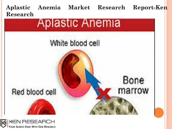
APLASTIC ANEMIA. Dr.Ramadas Nayak Professor & HOD Pathology Yenepoya Medical college Mangalore. APLASTIC ANEMIA. Hematopoietic stem cell (HSC) disorder characterized by: Pancytopenia (anemia, neutropenia and thrombocytopenia).

E N D
APLASTIC ANEMIA Dr.RamadasNayak Professor & HOD Pathology Yenepoya Medical college Mangalore
APLASTIC ANEMIA • Hematopoietic stem cell (HSC) disorder characterized by: • Pancytopenia (anemia, neutropenia and thrombocytopenia). • With markedly hypocellular bone marrow (less than 30% cellularity).
APLASTIC ANEMIA • Clinical Features • Any age of both sexes • Insidious • Progressive weakness, pallor and dyspnea due to anemia. • Frequent (mucocutaneous bacterial infections) or fatal infections due to neutropenia. • Bleeding manifestations in the form of petechiae, bruises and ecchymoses due to thrombocytopenia.
APLASTIC ANEMIA • Laboratory Findings • Peripheral Blood • Hemoglobin : Reduced • PCV: Reduced • Reticulocyte count: Markedly decreased.
APLASTIC ANEMIA • Peripheral smear: Pancytopenia, i.e. decreased red cells, neutrophils and platelets. • RBCs: Normocytic normochromic anemia– • WBCs: Total leukocyte count decreased. Neutrophils markedly diminished and neutropenia is a reflection of the severity of aplasia. • Initial stages, lymphocytes normal in number as the disease progresses their count decreases. • Platelets: Count is decreased.
Bone Marrow in aplastic anemia • Marrow aplasia—best appreciated in a bone marrow (trephine) biopsy • Cellularity: Marked hypocellularity. • Hematopoiesis: Paucity of all erythroid, myeloid and megakaryocytic precursors.– • Other cells: Lymphocytes and plasma cells are prominent.
No Splenomegaly • Diagnosis: Diagnosis is made with peripheral blood and bone marrow biopsy findings. • Prognosis: Unpredictable.
HEMOLYTIC ANEMIA • Definition • Hemolytic anemias are due to increase in the rate of red cell destruction (hemolysis).
HEREDITARY SPHEROCYTOSIS • Hereditary spherocytosis (HS) is a rare inherited hemolytic anemia resulting from the defect in the red cell membrane.
HEREDITARY SPHEROCYTOSIS • Etiopathogenesis • Autosomal dominant disorder • RBC membrane protein defect caused by various mutations. Most common mutations involve ankyrin, band 3, spectrin, or band protein 4.2.
HEREDITARY SPHEROCYTOSIS • Mechanism of Hemolysis in HS • Young HS RBCs are normal in shape. • But as they age, they undergo loss of membrane fragments in the circulation. • These small RBCs assume a spherical shape (spherocytes). • Spherocytesare rigid, inflexible and less deformable. • They get trapped in the spleen leading to premature destruction of spherocytes.
Laboratory Findings • Peripheral Blood • Hemoglobin: Decreased and level depends on degree of hemolysis. • Red cell indices: • MCV: Reduced (normal 82–98 fL) • MCHC: Raised and > 35 g/dL (normal 31–36 g/dL)
Peripheral smear • Very important for diagnosis • RBCs: • Spherocytesare most distinctive but not pathognomonic. • Spherocytesare small, dark-staining (hyperchromic) RBCs without any central pallor. • Polychromatophiliadue to reticulocytosis. • WBCs: Total leukocyte count (TLC) increased. • Platelets: Normal.
Osmotic Fragility Test • Osmotic fragility is increased and there is shift of the curve to the right
Clinical Features • Age: Anytime from the neonatal period to adulthood. • Family history: Most (75%) are inherited as autosomal dominant trait. • Anemia: Mild to moderate • Jaundice: Intermittent attacks, precipitated by pregnancy, fatigue, or infection. • Splenomegaly: Moderate (500 to 1000 g). • Gallstones: Pigment gallstones. • Aplastic crises: May be triggered by an acute parvovirus infection.