Download
1 / 37
441 likes | 702 Views
Aplastic Anemia. Dr Ramadas Nayak Professor & HOD Pathology Yenepoya Medical College Mangaluru. DEFINITION. Aplastic anemia is a chronic primary hematopoietic stem cell (HSC) disorder characterized by pancytopenia (anemia, neutropenia and thrombocytopenia)

E N D
Aplastic Anemia DrRamadasNayak Professor & HOD Pathology Yenepoya Medical College Mangaluru
DEFINITION • Aplastic anemia is a chronic primary hematopoietic stem cell (HSC) disorder characterized by • pancytopenia (anemia, neutropenia and thrombocytopenia) • associated with hypocellular bone marrow (less than 30% cellularity).
DEFINITION • Hematopoietic stem cells in the bone marrow are replaced by fat cells and result in deficient production of all the blood cells
ETIOLOGY • Aplastic anemia results from injury to hematopoietic stem cells in the bone marrow
Etiology- Acquired • Chemical agents • Certain drugs and agents (cancer chemotherapy drugs and the organic solvent benzene) cause marrow damage, which is dose related and reversible, when the offending agent is withdrawn.
Etiology- Acquired • Idiosyncratic: Some chemicals and drugs cause marrow suppression due to an idiosyncratic reaction to the drugs (e.g. chloramphenicol, phenylbutazone). • Physical agents: Whole-body irradiation can destroy hematopoietic stem cells. The damage is dose-dependent and follows either therapeutic irradiation or nuclear accidents.
Etiology- Acquired • Viral infections: Viral hepatitis of unknown types; CMV, HIV and EBV is associated in few cases with aplastic anemia. • Idiopathic: Most cases (approximately 65%) are idiopathic without any identifiable specific initiating etiological factor.
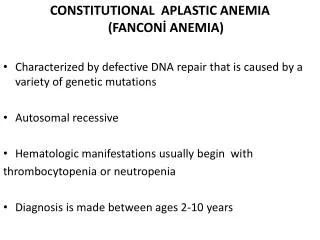
Etiology-Inherited • Fanconi anemia is a rare autosomal recessive inherited disorder. • Telomerase defects • Telomerase is required for cellular immortality and limitless replication of cells. • Hence, defects in telomerase activity can cause premature hematopoietic stem cell exhaustion and marrow aplasia
PATHOGENESIS • The exact pathogenesis of aplastic anemia is not known. • The mechanism of aplastic anemia falls under three main categories: • direct damage, • immune-mediated destruction of marrow progenitor cells and • primary stem cell abnormality
PATHOGENESIS • Direct damage to the hematopoietic stem cells and progenitor cells: • This may alter the ability of the cell to proliferate or differentiate. • The damage may be the result of known agent such as cytotoxic drugs, irradiation and viruses or unknown agent
PATHOGENESIS Immune-mediated destruction Acquired aplastic anemia is considered as an immune-mediated syndrome in which destruction of stem and progenitor cells occurs in the following ways: ––Chemical, viral or unidentified environmental antigens may produce acquired stem cell abnormality. Abnormal stem cells might express neoantigens and alter the stem cell antigenicity.
PATHOGENESIS Immune-mediated destruction ––Immunologically altered stem cell provokes a cellular immune response and activation of cytotoxic T lymphocyte. ––Activated TH1 cells produce cytokines such as interferon-γ (IFN-γ) and TNF. ––The cytokines suppress and destroy hematopoietic progenitors.
PATHOGENESIS Primary stem cell abnormality: Aplastic anemia may also be due to inherited defect in the stem cells.
CLINICAL FEATURES • Aplastic anemia can occur at any age of both sexes. • Insidious in onset and the initial presenting feature depends on the hematopoietic cell line predominantly affected. • Anemia: This causes progressive weakness, pallor and dyspnea. • Neutropenia: Presents as frequent (mucocutaneous bacterial infections) or fatal infections. • Thrombocytopenia: Results in bleeding manifestations in the form of petechiae, bruises and ecchymoses.
LABORATORY FINDINGS • Peripheral Blood • Blood and bone marrow picture is similar in idiopathic and secondary aplastic anemia. • ••Hemoglobin: Decreased. • ••PCV: Decreased. • ••Reticulocyte count: Reticulocyte count is markedly decreased (varies from 0.5 to 1%). It is known as reticulocytopenia and is a characteristic feature
LABORATORY FINDINGS • Peripheral smear: Shows pancytopenia, i.e. decreased red cells, neutrophils and platelets. In early stages, single cell line shows decreased count or bicytopenia. • ––RBCs: They are typically normocytic and normochromic, although slight macrocytosis may be occasionally present. • ––WBCs: Total leukocyte count is decreased. • Neutrophils are markedly diminished and neutropenia is a reflection of the severity of aplasia. • In the initial stages, lymphocytes are normal in number but with progression of the disease their count also decreases. • ––Platelets: Count is decreased.
LABORATORY FINDINGS • Bone Marrow • Marrow aplasia is best appreciated in a bone marrow (trephine) biopsy as the marrow involvement may be focal. • ••Cellularity: Marked hypocellularitywith replacement of more than 70% of the marrow cells by fat. • ••Hematopoiesis: Paucity of all erythroid, myeloid and megakaryocytic precursors. Initial stages may show focal areas of hematopoiesis. • ••Other cells: Lymphocytes and plasma cells are prominent.
LABORATORY FINDINGS • Splenomegaly is absent and in its presence the diagnosis of aplastic anemia should not be made. • Diagnosis: Diagnosis is made with peripheral blood and bone marrow biopsy findings.
Criteria for severe aplastic anemia (SAA) • Any two of the following for are required for the diagnosis: • 1. Granulocytes <0.5 × 10⁹/L • 2. Platelets <0.5 × 10⁹/L • 3. Reticulocytes <1% (corrected) • 4. Marrow cellularity <25%.
Differential diagnosis ofaplastic anemia • Aplastic anemia should be distinguished from other causes of pancytopenia such as “aleukemic” leukemia and myelodysplastic syndromes, which have similar clinical manifestations. • In aplastic anemia, the marrow is usually markedly hypocellular, with prominence of lymphocytes and plasma cells and • in contrast the myeloid neoplasms have abnormal myeloid progenitors.
Prognosis ofaplastic anemia • Prognosis of aplastic aplasia is unpredictable. • Withdrawal of toxic drugs can lead to recovery in some cases. • Spontaneous remission in idiopathic cases is unfortunately uncommon. • Bone marrow transplantation is the treatment of choice. • Immunosuppressive therapies will be helpful in older patients
Fanconi anemia • It is a rare inherited cause of aplastic anemia. • Peripheral blood in aplastic anemia shows pancytopenia with reticulocytopenia. • Bone marrow is markedly hypocellular with paucity of erythroid, myeloid and megakaryocytic precursors. • Marrow hypofunction is often associated with multiple congenital anomalies (hypoplasia of the kidney and spleen and bone anomalies).
Pancytopenia • The suffix (-penia) means lacking or a deficiency. • Definition: Combination of anemia, leukopenia and thrombocytopenia
Cytopenia- a deficiency in the production of one or more types of blood cells. • Leukopenia • Neutropenia • Thrombocytopenia • Osteopenia
Paroxysmal Nocturnal Hemoglobinuria • Paroxysmal nocturnal hemoglobinuria (PNH) is an acquired genetic defect in the cell membrane due to mutation in the hematopoietic stem cell. • Red cells in PNH are abnormally sensitive to complement-mediated intravascular hemolysis. • Severity of hemolysis is proportional to the number of these abnormal red cells. • It is the only hemolytic anemia caused by an acquired genetic defect
Main features of PNH • Intravascular hemolysis • Deficiency of GPI-linked proteins that regulate complement renders the red cells susceptible to complement-mediated hemolysis. • Chronic hemolysis without hemoglobinuria is more common than the paroxysmal and nocturnal hemolysis despite the name (nocturnal).
Main features of PNH • Intravascular hemolysis • Nocturnal hemolysis is due to reduced pH of blood during sleep which enhances the activity of complement. • Hemoglobin in acidic urine is converted into acid hematin which colors the urine dark brown. • The loss of heme iron in the urine (hemosiderinuria) eventually causes iron deficiency
Main features of PNH • Thrombosis • Absence of GPI-linked proteins in platelets and absorption of NO by free hemoglobin predisposes to thrombosis. • Thrombosis involving the hepatic, portal or cerebral veins is often the leading cause of death.
Summary • Aplastic anemia is characterized by reduction or cessation of production of blood cells. • Aplastic anemia may be acquired or congenital. The acquired aplastic anemia may be idiopathic or secondary to drugs, chemicals, radiation, infections, etc.
Summary • In acquired aplastic anemia the bone marrow aplasia is due to destruction of hematopoietic stem cells by direct or indirect damage. The indirect damage may result in expression of neoantigens and their subsequent destruction by immune mechanism. • The bone marrow aplasia is best appreciated in a bone marrow (trephine) biopsy. • Fanconianemia is a rare inherited cause of aplastic anemia.