Download
1 / 17
220 likes | 1.28k Views
Hematopoietic and lymphoreticular system. SYLLABUS: RBP( Robbins Basic Pathology ) Chapter : The Hematopoietic and Lyphoid Systems. Hematopoietic and lymphoreticular system. 51 Chronic Lymphocytic Leukemia (CLL)/ Small Lymphocytic Lymphoma (SLL) (lymph node) 116 Hodgkin Lymphoma (HL)

E N D
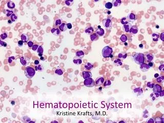
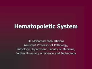
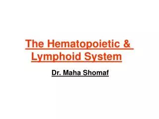
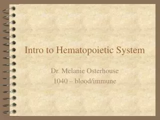
Hematopoietic and lymphoreticular system SYLLABUS: RBP(Robbins Basic Pathology) Chapter: TheHematopoietic and Lyphoid Systems
Hematopoietic and lymphoreticular system 51Chronic Lymphocytic Leukemia (CLL)/ Small Lymphocytic Lymphoma (SLL) (lymph node) 116Hodgkin Lymphoma (HL) 88Multiple myeloma 53Chronic Myelogenic Leukemia (CML) (liver) 26Amyloidosis (spleen)
Chronic Lymphocytic Leukemia (CLL)/ Small Lymphocytic Lymphoma (SLL) (lymph node) Lymph node architecture diffusely effaced by a predominant population of small lymphocytes containing round to slightly irregular nuclei with condensed chromatin and scant cytoplasm. These cells are mixed with variable numbers of larger cells called "prolymphocytes." In many cases, prolymphocytes gather together focally to form loose aggregates referred to as proliferation centers, so called because they contain relatively large numbers of mitotically active cells. When present, proliferation centers are pathognomonic for CLL/SLL.
Chronic Lymphocytic Leukemia (CLL)/ Small Lymphocytic Lymphoma (SLL) (lymph node)
Chronic Lymphocytic Leukemia (CLL)/ Small Lymphocytic Lymphoma (SLL) (lymph node)
Hodgkin Lymphoma (HL) Diagnostic Reed-Sternberg cells are large and have either multiple nuclei or a single nucleus with multiple nuclear lobes, each with a large inclusion-like nucleolus about the size of a small lymphocyte. The cytoplasm is abundant. Several variants of Reed-Sternberg cells are also recognized. Reed-Sternberg cells must be present in an appropriate background of non-neoplastic inflammatory cells (lymphocytes, plasma cells, eosinophils).
Multiple myeloma • Infiltrate of typical and/or atypical plasma cells. • some pleomorphism and binucleate forms • some cells more immature • may accumulate immunoglobulin in cytoplasm (Russell bodies) or nucleus (Dutcher bodies) • the anaplastic (plasmablastic, variant) type has a nucleus more like that of an immunoblast with a single large nucleolus
Chronic Myelogenic Leukemia (CML) (liver) • - marked leukocytosis of the peripheral blood • circulating cells are predominantly neutrophils, metamyelocytes, and myelocytes, with less than 10% myeloblasts • peripheral blood eosinophilia and basophilia are also common • up to 50% of patients have thrombocytosis early in the course of their disease
Amyloidosis (spleen) • Sago spleen pattern: • deposits largely limited to the splenic follicles, producing tapioca-like granules • - histologically the entire follicle may be replaced in advanced cases