Download
1 / 38
380 likes | 424 Views
Hematopoietic System. Clinical Pathology. Hematopoietic System. Blood supplies cells with water, nutrients, electrolytes, and hormone. Removes waste products Mainly CO 2 Cellular elements supply oxygen (RBC), protect against foreign organisms (WBC) and initiate coagulation (platelets).

E N D
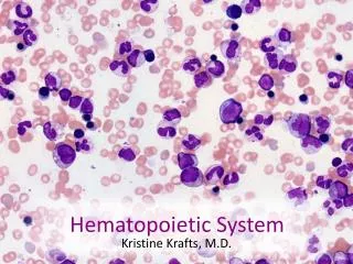
Hematopoietic System Clinical Pathology
Hematopoietic System • Blood supplies cells with water, nutrients, electrolytes, and hormone. • Removes waste products • Mainly CO2 • Cellular elements supply oxygen (RBC), protect against foreign organisms (WBC) and initiate coagulation (platelets). • Clotting factors
Hemoglobin • Normal values are usually 1/3 of the hematocrit. • Each hemoglobin molecule has 4 heme units attached to globulins. • Abnormal heme groups, cannot carry oxygen. • Carboxyhemaglobin- Hgb has a higher affinity for CO than O2. • Bright red blood • Methemoglobin- The Fe molecule is oxidized to Fe+3. • Blood becomes brown. • Tylenol toxicity in cats.
Oxygen-Hemoglobin Dissociation Curve • Increasing the temperature denatures the bond between hemoglobin and oxygen. • A decrease in pH causes a shift to the right causing more O2 to be released. • Organic phosphates decrease the affinity of hemoglobin to O2. • The purpose of the curve is to show the equilibrium of oxyhemoglobin at various partial pressures. • At high partial pressures of oxygen, usually in lungs, hemoglobin binds to oxygen.
Red Blood Cells • Function: • Carry oxygen to the tissues • Oxygen must be carried at enough pressure to permit rapid diffusion of oxygen. • The RBC is a vehicle for hemoglobin which is the carrier molecule for oxygen. • The sigmoid shape of curve is a result of the cooperative binding from the 4 hemoglobin molecules.
Transport of oxygen in the blood • 97% of oxygen transported from lungs to the tissues is carried in chemical combination with hemoglobin in RBC’s. • When the Po2, is high, O2 binds to hemoglobin. When Po2 is low, O2 is released.
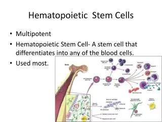
Erythropoiesis • Maturation of a RBC. • Only occurs in the bone marrow of normal adult animals. • Occurs in the spleen and liver of the fetus. • Maturation time usually takes 5 days. • Regulated by erythropoietin (EPO) which is increased in the presence of hypoxia. • In most species, the kidney is the sensor organ and major site of EPO.
Red Blood Cells (Erythrocytes) • No nucleus due to have to fold and squeeze through tight spaces. • Normocytes- cells look normal
Erythrocyte Life Span • Dog- 110 days • Cat- 70 days • Cow- 160 days • Horse- 145 days • Man- 120 days • Mouse- 30 days
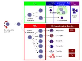
Erythrocyte Life Span • Stem Cell → Rubriblast → Prorubricyte → Metarubricyte → Rubricyte → Reticulocyte → RBC • Rubricyte- nucleated RBC releases in severe anemia. • No more mitotic division takes place after this stage. • One rubriblast may give rise to as many as 8-32 RBC’s.
Reticulocytes in the Peripheral Blood • Non-nucleated cell containing RNA which can be easily seen when stained with methylene blue. • Hallmark of erythrocyte regenerative response.
Polychromasia • Show a faint, bluish tint. These cells would appear to be reticulocytes if stained with methylene blue.
Nucleated Erythrocytes • Metarubricytes are sometimes found in the peripheral blood. • Usually observed with regenerative anemias. • May be found in non-regenerative states such as lead poisoning and hypoxia.
Macrocytosis • Erythrocytes are larger than normal. • Usually in the presence of regenerative anemia. • May be seen in FeLv infected cats. • Anisocytosis- increase variation of the size.
Microcytosis • Cells are smaller than normal which has been determined by the Mean Cell Volume (MCV). • A decreased MCV suggests that the cells are smaller than normal. • Usually occurs with iron deficiency cause by chronic blood loss or parasitism. • Haemobartonella • Feline Leukemia
Hypochromasia • RBC’s that have decreased density of the characteristic hemoglobin color. • Frequently observed in iron deficiency anemia caused by chronic blood los or parasitism.
Howell-Jolly Bodies • Nuclear remnants observed in young erythrocytes. • Often observed in cats and horses. • Can be seen in regenerative anemic animals. • Also may be seen with splenic disease or in an animal with the spleen removed.
Basophilic Stippling • Observed in RBC’s that contain abnormal aggregation of RNA. • Can be observed in cases of heavy metal poisoning with non-regenerative anemias or intense erythrogenesis in dogs, cats, and ruminants.
Spherocytes • Cells have a spheroid shape instead of the usual biconcave disk shape. • Have reduced cell membrane and are hypochromatic. • Seen most frequently in autoimmune hemolytic anemia (AIHA). • Usually seen in dogs.
Heinz Bodies • Particles of denature hemoglobin protien. • They stain with new methylene blue and appear as colorless bumps with quick stain. • May be caused by oxidant drugs and chemicals. • Normal cat blood may have 2-3%. • Spleen recognizes as abnormal and starts to lyse the cells.
Crenation • Identified as the presence of many irregular membrane projections involving most RBC’s. • It is usually an artifact due to slow drying of the blood film. • Commonly observed in pig blood but can be seen in any species.
Schistocytes • Also known as poikilocytes. • RBC’s with abnormal shape. • They are observed in fragmentation hemolysis caused by DIC, vascular neoplasia, endocarditis, and possibly iron deficiency anemia.
Target Cells and Folded Cells • Two types of leptocytes observed mainly in dogs. • Represent cells with an increases membrane-to-volume ratio not specific to any disease. • The cell membrane is thin and flimsy.
Rouleaux • RBC’s that in the form of stack of coins that has ben pushed over. • Commonly observed in horses and sometimes cats. • May indicate increase immunoglobulins in other species. • Autoimmune disease
Autoagglutination • Irregular clumping together of RBC’s caused by presence of anti-red cell antibody.
Normal Feline Blood • Feline RBC’s have less central pallor compared to canine blood. • Rouleaux and crenation are common features.
Normal Equine Blood • RBC’s have little central pallor and rouleaux is normal.
Normal Bovine Blood • RBC’s are often crenated. • Anisocytosis is sometimes observed in normal blood.
Platelets • Form the initial hemostatic plug whenever hemorrhage occurs. • The source of phospholipid which is needed for coagulation factors to interact to form a fibrin clot. • Produced in the bone marrow by megakaryocytes under the influence of thrombopoietin.
Laboratory Evaluation on Blood • CBC- Complete Blood Count • Involves: • Quantifying numbers of RBC’s , WBC’s, and platelets. • Making qualitative comments on cell morphology. • Determining the plasma protein level and color.
Blood Evaluation • Hematocrit- the % of RBC to plasma. Acquired by centrifuging blood in a microhematocrit tube for 5 minutes and then read. • Hemoglobin- 1/3 the hematocrit. • Red cell count- usually in the 5-15 x 106 ul range.
Instrumentation • Electronic cell counters- based on the principle that cells are poor electrical conductors. A measured volume of diluted blood is drawn between two electrodes, causing a resistance in an electrical current. • QBC- quantitative buffy coat system, utilizes differential centrifugation and quantification of cellular elements in a specialized microhematocrit tube.
Manual Procedures • Packed Cell Volume (PCV aka hematocrit)- % of RBC’s in the blood. • White Blood Cell Count- use of a hematocytometer and microscope involves observing the blood and counting the number of cells. • Evaluation of blood films- feathering technique of a drop of blood on a slide then staining before examining with a microscop.