Download
1 / 58
580 likes | 670 Views
Theme: Gene diseases of human L ecturer : ass. prof. T е tyana Bihunyak. The main questions: 1. Gene mutations 2. Gene diseases (metabolic disorders or molecular pathology) 2.1. Autosomal disorders 2.2. Sex-linked disorders 3. Indications for prenatal genetic counseling.

E N D
Theme: Gene diseases of human Lecturer: ass. prof. Tеtyana Bihunyak
The main questions:1.Gene mutations2. Gene diseases (metabolic disorders or molecular pathology)2.1. Autosomal disorders2.2. Sex-linked disorders3. Indications for prenatal genetic counseling
SingleGene disorders In 1966 about 1500 single gene disorders known. By 2012 over 6000 identified. There are between 50000-100000 structural genes so more disorders likely to be identified.
Single gene (monogenic or mendelian) disorders - 24/1000 live births (2.4%) 12 670 monogenic traits and diseases (incl. subtypes). For 9288 gene or its chromosomal location is known. 971 with a known molecular defect 1. Autosomal dominant disorders - 15/1000 Examples: Familial hypercholesterolaemia, brachydactylia 2. Autosomal recessive disorders – 7.5/1000 Examples: Cystic fibrosis, phenylketonuria, hydrocephalus,albinism 3. X-linked disorders (dominant and recessive) - 1.5/1000 Examples: haemophylia, hypophosphataemia 4. Holandric disorders
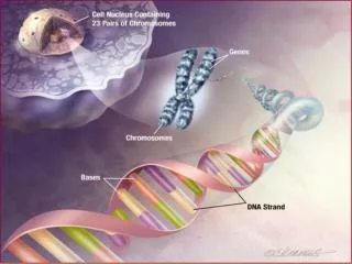
Gene disorders(metabolic disorders) or molecular pathologyare caused by mutation in the genesA gene mutation or point mutation (since it applies to a particular gene locus) is the result of a change in the nucleotide sequence of the DNA molecule in a particular region of the chromosome
These mutations take the form of the duplication, insertion, deletion, inversion or substitution of bases. Such a change in the base sequence of the gene is transmitted to mRNA during transcription and may result in a change in the amino acid sequence of the polypeptide chain (modified polypeptide) produced from it during translation at the ribosomes
Gene mutations occurring during gamete formation are transmitted to all the cells of the offspring and may be significant for the future of the species. Somatic gene mutations which arise in the organism are inherited only by those cells derived from the mutant cells by mitosis.
Task In some regions of South Africa there is a spread sickle-shaped cell anemia, in which erythrocytes have shape of a sickle as a result of substitution of glutamine by valine in the hemoglobin molecule. What is the cause of this disease? A. Transduction • Gene mutation • Genomic mutations D. Crossin-over E. Disturbance of mechanisms of genetic information realisation
Autosomal-Dominant inheritance: • 1) a trait can effect both sexes (female and male can be ill); • 2) the trait is inherited vertically in the pedigree (it affects every generation); • 3) one or both parents of ill child are ill
Autosomal dominant disorders N Mating types AAAa aa Risk for progeny 1.*Aa x aa 0 1/2 1/2 1/2 2. Aa x Aa 1/4 1/2 1/4 3/4 3. AA x aa 0 1 0 1 4. AA x Aa 1/2 1/2 0 1 5. AA x AA 1 0 0 1 Affected individuals are underlined. * - the most often mating type
Autosomal dominant disorders. Pedigree. I II III IV 1 2 1 2 3 4 5 6 7 8 1 2 3 4 6 8 9 10 11 12 5 7 6 1 2 3 4 5
Familial hypercholesterolaemia (FH) FH accounts for 5% of the coronary artery disease (CAD) seen in the Western world Heterozygotes (Aa) 1:500 Homozygotes (AA) 1:1.000.000 The most frequent type of monogenic hypercholesterolaemia Mutation in the gene coding for the low-density lipoprotein(LDL) receptor The LDL receptor gene maps to chromosome 19p13 and is made up of 18 exons The mature mRNA codes for a protein of 839 amino acids
Diagnosis: 1. Clinical symptoms: premature atherosclerosis, xanthomatosis (fatty deposits occur in various parts of the body). Variability in the age of onset (majority - in third or fourth decade). 2. Family history. 3. Biochemical findings Increased fasting total cholesterol. 4. DNA analysis.
Achondroplasia. Autosomal dominant trait, occur with a frequency of 20/1.000.000. Affected persons are characterized by a peculiar form of dwarfism with short limbs, an enlarged head, small face and depressed nasal bridge. The trunk is normal in size, but lordosis occurs
null mutation dominant negative mutation TYPICAL TYPE II OI TYPICAL TYPE I OI NORMAL a1 2n n n normal chains + n mutant chains a2 n / 4 procollagen molecules PROCOLLAGEN n / 2 procollagen molecules n procollagen molecules DEGRADED Collagen gene mutation
Prognathism is the positional relationship of the mandible to the skeletal base where the jaw protrudes beyond a predetermined imaginary line in the coronal plane of the skull. The word prognathism derives from Greek pro (forward) and gnathos (jaw).An individual's top teeth and lower teeth do not align properly. It is incompletely penetrant autosomal dominant trait
Progeria, or early aging, is a rare autosomal dominant disorder that apparently arises through new mutations. Children affected by this disorder start to show signs of advanced aging when they are only five or six. Their skin wrinkles, their hair thins, they start suffer arthritis, and their blood vessels show arteriosclerosis. Frequently, affected youngsters die of heart disease before they are 10 years old
Autosomal-Recessive inheritance: • 1) a trait can effect both sexes; • 2) the trait is inherited horizontallyin the pedigree (it does not affect every generation); • 3) parents of ill child can be healthy in the phenotype, but theyare heterozygous (individuals who have affected children must both be carriers)
Autosomal recessive disorders N Mating types AA Aa aa Risk for progeny 1.* Aa x Aa 1/4 1/2 1/4 1/4 2. Aa x AA 1/2 1/2 0 0 3. AA x aa 0 1 0 0 4. Aa x aa0 1/2 1/2 1/2 5. aa x aa 0 0 1 1 Affected individuals are underlined. * - the most common mating type
Pedigree of autosomal recessive disorder I II III IV Affected Consanguineous mating
Parental consanguinity (%) in AR pathology Disease % Cystic fibrosis in Nothern Europe1 – 2 Albinism 5 Phenylketonuria12.5 Tay-Sachs disease 27 Alkaptonuria 60
is an inherited error of metabolism caused by a deficiency in the enzyme phenylalanine hydroxylase. • Loss of this enzyme results in organ damage, unusual posture. It characterized by mental retardation, hypopigmentation of hair and skin, and mousy odor. • PKU is an autosomal recessive disorder, caused by mutations in both alleles of the gene for phenylalanine hydroxylase (PAH), found on chromosome 12. • Diagnosis. Screening tests for all babies • Treatment. Phenylalanine-low diet. Low protein foods such as fruits, vegetables, and some cereals are may be allowed. Phenylketonuria (PKU)
Albinism • is an autosomal recessive disorder • Lack of dark pigment melanin in the skin, hair and eye • It is caused by the absence of the enzyme tyrosinase, which is necessary for the synthesis of melanine from typosine
Hydrocephalus an abnormal increase in the amount of cerebrospinal fluid within the ventricles of the brain.Hydrocephalus makes the head enlarge. It cased by obstruction to the outflow of cerebrospinal fluid from the ventricles
Alkaptonuria • Is an autosomal recessive disorder • defect in theenzyme homogentisate 1,2-dioxygenase, which participates inthe degradation oftyrosine. • Homogentisic acidand its oxide,calledalkapton,accumulate in the blood and are excreted in urine in large amounts (hence -uria). • Pigmented the sclera of the eyes(often only at a later age); the skin darkened in sun-exposed areas; urine may turn brown if collected and left exposed to open air; kidney stones;osteoarthritis and intervertebral discs calcification • Both blood plasma and urine can be used for diagnosis (chromatography).
Tay-Sachs disease • is an autosomal recessive disorder • results from mutations in the HEXA gene on • human chromosome 15 • gangliosidesaccumulate in the brain's nerve cells, • leading to the premature death of the cells • It characterized by infantile onset (3-6 months), • doll-like facies, cherry-red macular spot, • early blindness, deafness • Death usually occurs before the age of four • There is no known cure or treatment
Sickle cell anaemia in humans is an example of base substitution mutation affecting a base in one of the genes involved in the production of haemoglobin
H N 2 O H C C H H 3 3 C H C 2 C O C H 2 H H N 2 C O H H N O H 2 O H Valine Glutamine Hemoglobin and Sickle Cell Anemia • Single base mutation in DNA • A to T transversion • Single amino acid change in the protein • Glutamine to Valine
Sickling Cells Polymers of hemoglobindeform red blood cells Normal Sickle
Sickle Cell Anemia • Recessive trait • Symptoms: • Chronic hemolytic anemia • Severe pain • Rapid septicemia (infection) • Asplenia (no spleen left)
How Was the Mutation Selected? • Malaria • Mosquito born plasmodium parasite • Some sickling is good • Heterozygotes have the advantage!
Task Gene disease with characteristics: dwarfism, large head, short limbs and trunk, life span is normal: A. Marfan Syndrome. B. Achondroplasia. C. Albinism. D. Phenylketonuria. E. Sicle-cell anaemia.
Idiogram of human X chromosome Pseudoautosomal region 1 (PAR 1) Steroid sulphatase Kallmann’s syndrome Duchenne’s muscular dystrophy Becker’s muscular dystrophy Dystrophin X-inactivation center (XIST) HGPRT Hemophilia B G6PD, Hemophilia A Pseudoautosomal region 2 (PAR 2)
Human X-recessive traits (in 10.000 males) 1.Red-green colour-blindness 800 Normal colour vision depends upon the products of three loci – blue (BCP) on chr.7 and red (RCP) and green (GCP) in Xq28 2.Non-specific X-linked mental retardation 5 3.Duchenne muscular dystrophy 3 4.Becker muscular dystrophy 0.5
5. Haemophilia A (factor VIII deficiency) 2 Xq28. Recurrent haemorrhage postoperatively and spontaneously into soft tissues and joints 6. Haemophilia B (factor IX deficiency) 0.3 s. Christmas disease Xq27.1 7. X-linked ichthyosis2 8. X-linked agammglobulinaemia 0.1
X-Linked Recessive Inheritance • Males show disorder more than females • Son cannot inherit disorder from his father
Hemophilia. Hemarthroses (extravasation of blood into a joint or its synovial cavity) It is caused by defect ofthe blood which prevents its clotting due to deficiency of coagulation factors VIII (hemophilia A)or IX (hemophilia B).
Task A normal woman whose father had hemophilia A marries a man who also has hemophilia A. What is the chance their son will have the disorder? A. 100 % B. 50 % C. 75 % D. 25 % E. 0 %
Duchenne muscular dystrophy (DMD) • Becker muscular dystrophy (BMD) X – recessive disorder A French neurologist Duchenne described a case in 1861. Becker muscular dystrophy (BMD) – a similar but milder condition. Due to mutations in the same gene. Frequency DMD 1: 3.500 BMD 1:20.000 Gene located in the X-chromosome –Xp21. The DMD gene is one of the largest yet identified in man. The gene contains at least 79 exons and is expressed in muscle and in neurones of the cerebral cortex.
The gene product – protein dystrophin. Dystrophin is located at or close to the muscle membrane where it is thought to act as a link between extracellular laminin and intracellular actin. 1/3– new mutations, 2/3 – inherited cases (mother-heterozygote). DMD – deletions (2/3 cases) with a frameshift or nonsense mutations. No synthesis of dystrophin or very reduced. BMD – 5-10% of normal dystrophin quantity. Deletion without a frameshift or missense mutations. Diagnosis - symptomatology, pedigree analysis, elevated creatine kinase activity in serum, muscle biopsy, electromyography, DNA diagnosis (incl. prenatal DNA diagnosis). Identification of heterozygotes – elevated creatine kinase activity in serum (reveals 75% of Hz), pedigree analysis, DNA analysis.
Schematic representation of the probable structure of the dystrophin protein molecule which is depicted as a dimer linking intra-cellular actin with extra-cellular laminin Extracellular laminin 6 glycopro- tein complex Muscle membrane Dystrophin dimer Intracellular actin
X-Linked Dominant inheritance: 1) a trait affects mostly females; 2) an affected male passes the trait to all his daughters
X-linked dominant disorders Nr. Mating types XAXA XAXa XaXa XAY XaY Risk for progeny 1.* XAXa x XaY 0 1/4 1/4 1/4 1/4 1/2 2. XAXA x XaY 0 1/2 0 1/2 0 1 3.* XaXa x XAY 0 1/2 0 0 1/2 1/2 4. XAXa x XAY 1/4 1/4 0 1/4 1/4 3/4 5. XAXA x XAY 1/2 0 0 1/2 0 1 Affected individuals are underlined * - the most common mating types
Human X- dominant diseases Vitamin D resistant rickets s. Familial hypophosphataemic rickets s. Hypophosphataemia. 1 / 20 000. Failure of the intestinal epithelium to transport P. Growth retardation.Childhood rickets. Reduced serum phosphate. Gingival sinuses. Delayed eruption of teeth. Skull abnormalities. Both primary and secondary teeth involved. High pulp horns.
Hypophosphatemia (Vitamin D-resistant rickets) Not enough calcium salts are deposited in the bones to make them rigid: consequently they become soft and malformed
Enamel hypoplasiais hereditary defect that cause holes and cracks to appear around the crowns of the teeth. It is inherited as X-linked dominant trait