Download
1 / 3
30 likes | 171 Views
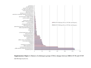
a. b. #1. #2. #3. #5 Patient number. #4. BEAS-2B. Sq Ad Ad Ad Ad Squamous/Adeno. c. H460. Caspase 8 gene expression. N T N T N T N T N T N ormal/ T umour. DR5. DR4. β -Actin. #11. #12. #13. #15 Patient number. #14.

E N D
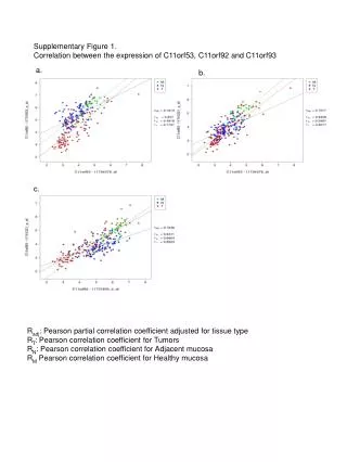
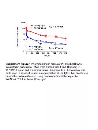
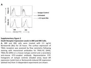
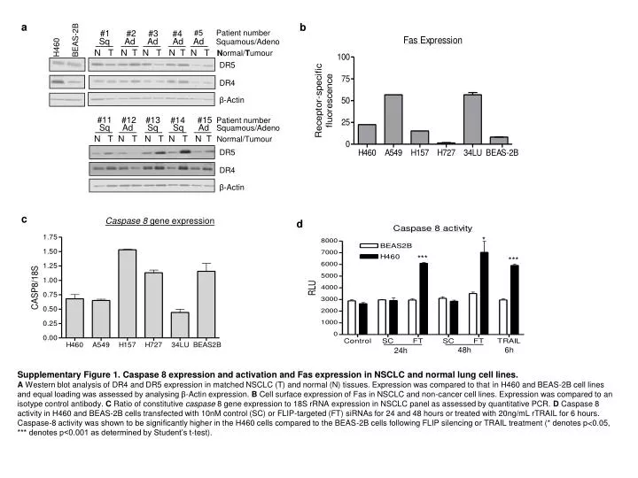
a b #1 #2 #3 #5 Patient number #4 BEAS-2B Sq Ad Ad Ad Ad Squamous/Adeno c H460 Caspase 8 gene expression N T N T N T N T N T Normal/Tumour DR5 DR4 β-Actin #11 #12 #13 #15 Patient number #14 Sq Ad Sq Sq Ad Squamous/Adeno N T N T N T N T N T Normal/Tumour DR5 DR4 β-Actin d * *** *** 48h 6h 24h Supplementary Figure 1. Caspase 8 expression and activation and Fas expression in NSCLC and normal lung cell lines. A Western blot analysis of DR4 and DR5 expression in matched NSCLC (T) and normal (N) tissues. Expression was compared to that in H460 and BEAS-2B cell lines and equal loading was assessed by analysing β-Actin expression. B Cell surface expression of Fas in NSCLC and non-cancer cell lines. Expression was compared to an isotype control antibody. C Ratio of constitutive caspase 8 gene expression to 18S rRNA expression in NSCLC panel as assessed by quantitative PCR. D Caspase 8 activity in H460 and BEAS-2B cells transfected with 10nM control (SC) or FLIP-targeted (FT) siRNAs for 24 and 48 hours or treated with 20ng/mL rTRAIL for 6 hours. Caspase-8 activity was shown to be significantly higher in the H460 cells compared to the BEAS-2B cells following FLIP silencing or TRAIL treatment (* denotes p<0.05, *** denotes p<0.001 as determined by Student’s t-test).
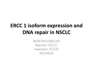
a c e f H460 d b H460 SC DR4 DR5 siRNA (10nM) DR4 *** β-Actin *** *** *** *** DR5 5µM cis β-Actin FT SC Supplementary Figure 2. Role of death receptors in regulating TRAIL and FLIP siRNA induced cell death in NSCLC. A Cell surface expression of DR4, DR5 and Fas in H460 cells transfected with 10nM control (SC), DR4, DR5 or Fas targeted siRNAs for 24 hours. Expression was compared to an isotype control antibody. B Western blot analysis showing the expression of DR4 and DR5 in H460 cells transfected with 10nM SC, DR4 or DR5 targeting siRNA for 24 hours. Equal loading was confirmed by analysing β-Tubulin expression. C Flow cytometry analysis of apoptosis in H460 cells treated with rTRIAL and co-treated with a TRAIL neutralising antibody (anti-TRAIL) for 24 hours. Apoptosis was assessed by analysing the sub-G0/G1 fraction of cells. The TRIAL neutralising antibody was found to significantly reduce TRAIL-induced apoptosis in H460 cells. D Flow cytometry analysis of apoptosis in H460 cells transfected with 10nM SC, DR4, DR5 and Caspase-8 (C8) siRNA for 24 hours followed by treatment with 5ng/mL TRAIL for 24 hours. Apoptosis was assessed by analysing the sub-G0/G1 fraction of cells. DR4+5 and C8 siRNAs were found to significantly rescue TRAIL-induced apoptosis (*** denotes p,0.001, Student’s t-test). E Flow cytometry analysis of apoptosis in H460 cells transfected with 10nM DR4 and/or DR5 siRNA for 24 hours followed by transfection with 1nM SC or c-FLIP targeted (FT) siRNA for 24 hours. Cells were co-treated with 2.5ng/mL TRAIL for the final 24 hours. F Flow cytometry analysis of apoptosis in H460 cells transfected with 10nM DR4 and DR5 siRNA for 24 hours followed by transfection with 1nM SC or c-FLIP targeted (FT) siRNA for 24 hours. Cells were co-treated with 5µM cisplatin for 48 hours. Apoptosis was assessed by analysing the sub-G0/G1 fraction of cells. DR4+5 siRNA was found to significantly inhibit apoptosis induced by TRAIL, FT siRNA + TRAIL, FT siRNA and FT siRNA + cisplatin (*** denotes p,0.001, Student’s t-test).
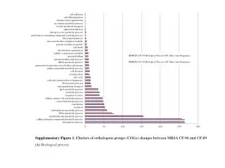
a b *** *** *** *** Supplementary Figure 3. Effect of FLIP silencing on taxol-induced apoptosis in NSCLC and normal lung cell lines. A Flow cytometry analysis of apoptosis in NSCLC cells transfected with 1nM non-silencing control (SC) or FLIP-targeted (FT) siRNAs for 48h. Transfected cells were co-treated with taxol as indicated. The combined effects of FT siRNA and taxol were shown to be supra-additive (*** denotes p<0.001 as determined by two-way ANOVA). B Flow cytometry analysis of apoptosis in BEAS-2B and 34LU cells transfected with 1nM SC or FT siRNA for 48h. Transfected cells were co-treated with taxol as indicated. Apoptosis was assessed by analysing the sub-G0/G1 fraction of cells. The normal lung cell line models were not sensitized to taxol following FLIP silencing.