Download
1 / 39
820 likes | 2.1k Views
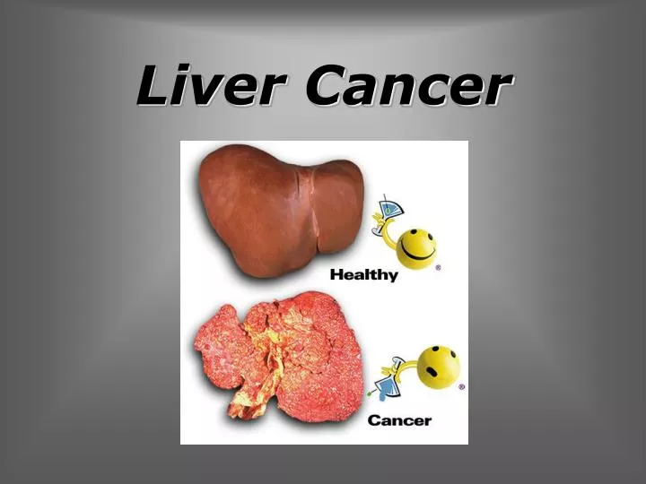
Liver Cancer. Liver Cancer. Hepatocellular carcinoma is the sixth most common cancer worldwide in terms of numbers of cases. It is the third most common cause of death from cancer (third to lung and gastric cancer).

E N D
Liver Cancer Hepatocellular carcinoma is the sixth most common cancer worldwide in terms of numbers of cases. It is the third most common cause of death from cancer (third to lung and gastric cancer). The remaining malignant tumors are fibrolamellar carcinoma, intrahepatic cholangiocarcinoma (10% to 25% of liver cancers), cystadenocarcinoma, angiosarcoma, hepatoblastoma, and undifferentiated embryonal sarcoma. Worldwide, the major risk factors for liver cancer are infection with the hepatitis B and C viruses. More than 75% of cases worldwide, and 85% of cases in developing countries, are caused by these two viruses. From: Global Cancer Statistics, 2002 -- Parkin et al_ 55 (2) 74 -- CA A Cancer Journal for Clinicians
Liver Cancer Frequency: • 82% of cases (and deaths) are in developing countries (55% in China alone). • The areas of high incidence are sub-Saharan Africa, eastern and southeastern Asia, and Melanesia. • The incidence is low in developed areas, Latin America, and southcentral Asia. From: Global Cancer Statistics, 2002 -- Parkin et al_ 55 (2) 74 -- CA A Cancer Journal for Clinicians
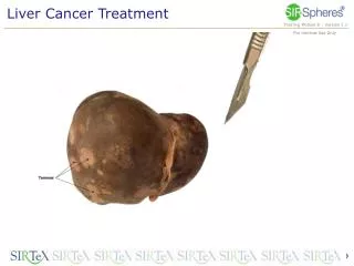
Liver Cancer • HCC is a malignant tumor of hepatocellular origin. • Grossly, HCC can undergo hemorrhage and necrosis because of a lack of fibrous stroma. • There are 3 growth patterns of HCC: • Solitary mass - Often large • Multifocal or nodular pattern - Multiple nodules • Diffuse - Multiple, small foci scattered diffusely throughout the liver
Liver Cancer • Clinical presentation • varies among high-incidence and low-incidence regions: • In high-incidence regions (ie, Asia, Africa), clinical presentation of HCC tends to be aggressive and includes bleeding, hepatic rupture, and hemoperitoneum. 2. In low-incidence regions (ie, Western Hemisphere), clinical presentation of HCC tends to be less aggressive and includes symptoms such as fever of unknown origin,abdominal pain, malaise, weight loss, and hepatomegaly. Jaundice is rare. In high-incidence regions of the world, the male-to-female ratio is approximately 8:1. In low-incidence regions, the male-to-female ratio is approximately 2:1. In high-incidence regions of the world, patients present at age 30-50 years. In low-incidence regions, patients present at age 70-80 years. (Patients with cirrhosis may present earlier)
Liver Cancer Preferred Examination: • Imaging studies are an important part of diagnosing HCC, but which imaging procedure is used differs in the United States versus the rest of the world. • Most studies screening for HCC have been performed outside the United States where ultrasound combined with AFP measurement achieves greater sensitivity and specificity than other methods. • In contrast, in a recent study of United States patients with advanced cirrhosis, spiral CT was found to have an 88% sensitivity for detecting HCC, which was significantly better than the 59% sensitivity of abdominal US. • Fine-needle biopsy of the mass should always be considered when the diagnosis is unclear, but theoretically, it carries risks for percutaneous needle tract seeding of tumor and bleeding. From: Hepatocellular carcinoma. A concise guide to its status and managementScott C. Ulmer, MD; POSTGRADUATE MEDICINEVOL 107 / NO 5 / 2000
Liver Cancer Preferred Examination: Radiologic evidence of cirrhosis, vascular invasion, or multifocal disease is typical in hepatocellular carcinoma.
Liver Cancer Ultrasound • US appearance of HCC is variable. • The quality of a US examination is operator dependent.
Liver Cancer Ultrasound Vascular invasion Liver US: large HCC with involvement of right hepatic vein (white arrows: hepatic veins)
Liver Cancer Ultrasound The 3-D US is useful in the demonstration of continuity of the intranodular vessels. This application provides better understanding of the hemodynamic vasculature in a tumor
Liver Cancer CT Findings: • CT appearance of HCC varies depending on tumor size and the imaging phase. • Unenhanced CT typically reveals an iso-hypodense mass. If the mass is large, central areas of necrosis may be seen. • In the hepatic-arterial phase, lesions typically are hyperdense (relative to hepatic parenchyma) as a result of hepatic-arterial supply. Larger tumors may have necrotic central regions that typically are hypodense. • In the portal-venous phase, small lesions may be isodense or hypodense and difficult to see, since the remainder of the liver increases in attenuation. Larger lesions with necrotic regions remain hypodense. • In the delayed-postcontrast phase, small lesions may be inconspicuous on late phases. Delayed phase scans may show a tumor capsule, one of the more specific signs indicating HCC.
Liver Cancer CT Findings: Micro - HCC (a) precontrast CT scan demonstrates a hypoattenuating mass (b) arterial-phase spiral CT scan showed the mass as a hypervascular area (c) portal-phase CT scan demonstrate a hypoattenuating mass.
Liver Cancer CT Findings: Small HCC, with a typical finding and washout. Transverse hepatic arterial phase CT image of midliver. The mass enhances fairly homogeneously but much less than the aorta. Portal venous phase image at the same level. The mass is now hypoattenuating to the liver and blood pool. From:Discrimination of Small Hepatic Hemangiomas from Hypervascular Malignant Tumors …Tonsok Kim et al.Radiology. 2001;219:699-706
Liver Cancer CT Findings: Unenhanced and contrast-enhanced axial scans: In the unenhanced upper images a huge, mildly, inhomogenously attenuating mass can be seen in the right lobe of the liver, which enhances inhomogenously (in the lower two pictures).
Liver Cancer CT Findings: Solitary HCC (a) Transverse nonenhanced CT scan shows a large mass (arrows) in the left lobe that is hypoattenuating to the liver. (b) Transverse CT scan obtained during the hepatic arterial phase after bolus injection of contrast material demonstrates heterogeneous enhancement of the tumor (arrows). (c) Transverse CT scan obtained during the portal venous phase demonstrates the heterogeneous tumor and thrombosis of the left portal vein (arrow). From: Hepatocellular Carcinoma in Noncirrhotic Liver: CT, Clinical, and Pathologic Findings in 39 U.S. ResidentsGiuseppe Brancatelli et al. Radiology 2002;222:89-94
Liver Cancer CT Findings: Multifocal HCC (a) Transverse nonenhanced CT scan shows multiple, hyperattenuating lesions (solid arrows) with a central hypoattenuating, necrotic portion (open arrow), well seen in the largest lesion. (b) Transverse CT scan obtained during the hepatic arterial phase after bolus injection of contrast material shows hyperattenuation of the lesions (arrows). The central portions remain hypoattenuating. (c) Transverse CT scan obtained during the portal venous phase shows that the mass is isoattenuating to the liver parenchyma. Note the capsule (arrows) around the largest lesion. From: Hepatocellular Carcinoma in Noncirrhotic Liver: CT, Clinical, and Pathologic Findings in 39 U.S. ResidentsGiuseppe Brancatelli et al. Radiology 2002;222:89-94
Liver Cancer MRI Findings: MRI is less sensitive than angiographically assisted helical CT in diagnosing HCC and is currently used to further characterize the disease within a nodular liver. • HCC on T1-weighted images may be isointense, hypointense, or hyperintense relative to the liver. • On T2-weighted images, HCC usually is hyperintense. • Gadolinium-enhanced MRI typically demonstrates that HCCs enhance, usually in the arterial phase and particularly if they are small. • Pre- and postcontrast MRI has a 70-85% chance of detecting a solitary mass of HCC.
Liver Cancer MRI Findings: MR imaging appearance of an HCC with a tumor capsule in a noncirrhotic liver. (a) Axial fat-saturated T2-weighted fast SE image shows an HCC with predominantly high signal intensity (arrow). Owing to its fibrotic nature, the tumor capsule has low signal intensity on T2-weighted images and therefore is not visible. (b) Axial gadolinium-enhanced 3D GRE image obtained during the arterial phase shows intense, nearly homogeneous enhancement of the lesion (arrow). This appearance may simulate FNH. (c) Axial gadolinium-enhanced 3D GRE image obtained during the delayed phase shows enhancement of the tumor capsule surrounding the lesion (arrow), which demonstrates complete washout. From: Focal Nodular Hyperplasia: Findings …Shahid M. et al. Radiographics. 2004;24:3-17
Liver Cancer MRI Findings: MR imaging appearance of an HCC with a central scar: (a) Axial fat-saturated T2-weighted fast SE image shows a predominantly high-signal-intensity lesion with a low-signal-intensity central scar. (b) Axial gadolinium-enhanced fat-saturated 2D T1-weighted GRE image obtained during the delayed phase shows washout of contrast material in most of the lesion and an enhanced tumor capsule. The central scar remains mainly unenhanced. From: Focal Nodular Hyperplasia: Findings …Shahid M. et al. Radiographics. 2004;24:3-17
Liver Cancer • Screening for Liver Tumors • Serum alpha-fetoprotein (AFP)levels are elevated in many patients with HCC. However, AFP measurement is not an ideal test because it lacks both sensitivity and specificity. • Real-time US is the most cost-effective modality for evaluating patients with HCC. • Modifications in CT have increased its sensitivity in patients with HCC from 50% or 60% to more than 90%. • In North America, screening for HCC in patients with HCV infection is widely accepted by hepatologists. In general, patients are screened by AFP measurements and USevery 6 to 12 months. • In patients with equivocal findings on CT, MRI is useful for its ability to accurately identify fatty infiltration and small HCCs A) CT scan demonstrates no focal liverlesion. B) Gadolinium DTPA-enhanced MRI reveals multiple focal enhancing liver lesions consistent with multifocal HCC (arrows).
Liver Cancer From:Flickinger et al
Metastatic Liver Tumors • The liver is the second most commonly involved organ by metastatic disease, after the lymph nodes. • In Europe and the United States, a focal liver lesion is more likely to represent a metastatic deposit than a primary malignancy. • The liver may be the site of metastasis from virtually any primary malignant neoplasm, but the most common primary sites are the eye, colon, stomach, pancreas, breast, and lung. • In children, the most common liver • metastases are from a neuroblastoma, • a Wilms tumor, or leukemia.
Metastatic Liver Tumors • Most liver metastases are multiple, involving both lobes in 77% patients, and only 10% are solitary. • Multiple tumors often vary in size. • Growing metastases compress adjacent liver parenchyma, causing atrophy and forming a connective tissue rim. • Large metastases often outgrow their blood supply, causing hypoxia and necrosis at the center of the lesion.
Metastatic Liver Tumors CT is the examination of choice for evaluating liver metastases. • Metastases may appear in a multitude of ways on CT scans. • The majority of liver metastases are hypovascular (hypoattenuating) compared with surrounding parenchyma Two rim enhancing metastases of lung cancer are present in the liver in contrast enhanced CT scan. CT scan with contrast - metastases from a colonic adenocarcinoma.
Metastatic Liver Tumors • During portal venous scanning, the attenuation of the normal liver parenchyma increases, revealing the relatively hypoattenuating metastases CT examination: Postcontrast axial scans: The focal lesions are more prominent due to contrast-enhancement of the normal parenchyma. CT examination: Unenhanced axial scans: Numerous, mostly round-shaped hypodens lesions of different size are visible in both lobes of the liver.
Metastatic Liver Tumors • Hyperattenuating lesions due to increased tumor vascularity are uncommon. • On arterial phase enhanced scans, these vascular metastases show homogenous enhancement compared with the surrounding liver Arterial phase CT scan- metastases from a brest cancer
Metastatic Liver Tumors 48-year-old woman with hypervascular hepatic metastases (neuroendocrine primary tumor). Arterial phase CT scan shows multiple hyperenhancing masses in liver. Hypervascular masses in patients with known hypervascular primary tumors (such as renal cell carcinomas, pancreatic islet cell tumors, pheochromocytomas, melanomas, and breast carcinomas)should be regarded as metastasis until proven otherwise. From: CT of Focal Nodular Hyperplasia of the LiverStephanie K et al.AJR 2000; 174:705-712
Metastatic Liver Tumors Ultrasound • The US appearance of liver metastases is nonspecific. • However, the presence of multiple hepatic nodules of different sizes within the liver is nearly always due to metastases. • The echogenicity is dependent on tumor vascularity; the cellular composition; the degree of tissue invasion; and the presence or absence of necrosis, fibrosis and fatty change. Multiple metastases in the liver.
Metastatic Liver Tumors Growing metastases compress adjacent liver parenchyma, causing atrophy and forming a connective tissue rim.
Metastatic Liver Tumors Large metastases often outgrow their blood supply, causing hypoxia and necrosis at the center of the lesion.
Metastatic Liver Tumors Approximately one half the patients with liver metastases have clinical signs of hepatomegaly or ascites
Metastatic Liver Tumors MRI Findings • Liver metastases have a variety of appearances on MRI. • Most liver tumors benign or malignant appear as hypointense lesions on T1-weighted images and hyperintense lesions on T2-weighted images. • (There are a few exceptions to this rule, for example, metastatic melanoma, which exhibits high signal intensity on T1-weighted MRIs relative to the liver) • Heavily T2-weighted images are useful in differentiating hemangiomas and cysts because the signal intensity is higher in these benign lesions as compared with liver metastases.
Metastatic Liver Tumors The patient has known colon carcinoma with liver metastases. T1 (left) and (right) T2 axial images of the liver.
Metastatic Liver Tumors Hypervascular metastases from carcinoid tumor 18’’ 90’’ • Axial T2-weighted image shows multiple areas of moderate hyperintensity. • Unenhanced axial T1-weighted image shows corresponding areas of moderate hypointensity (arrowheads). • Axial T1-weighted image obtained 18 sec after initiation of IV contrast administration shows corresponding areas of intense, homogeneous, or peripheral ring enhancement of lesions. • Axial T1-weighted image obtained 90 sec after initiation of IV contrast administration shows peripheral washout (arrow), a feature often apparent in hypervascular metastases. From: Spectrum of MRI Appearances of Untreated Metastases of the LiverIoana-Maria IM et al. AJR 2003; 181:809-817
Metastatic Liver Tumors • Morphologic characteristics on T2-weighted images that suggest metastatic liver disease include the following: • heterogeneous signal intensity with irregular and indistinct outer margins • smooth or irregular central area of high signal intensity with a surrounding ring of signal intensity lower than that of the central focus but higher than that of the adjacent normal liver.
Metastatic Liver Tumors Hypovascular metastasis from colon adenocarcinoma 18’’ 90’’ • Axial T2-weighted image shows large lesion of heterogeneous signal intensity with central areas of marked hyperintensity. • Unenhanced axial T1-weighted image shows lesion as area of moderate hypointensity. • Axial T1-weighted image obtained 18 and 90 sec after initiation of IV contrast administration shows thin peripheral ring enhancement From: Spectrum of MRI Appearances of Untreated Metastases of the LiverIoana-Maria IM et al. AJR 2003; 181:809-817
Metastatic Liver Tumors Nuclear medicine techniques • Hepatic arterial perfusion scintigraphy • (colorectal cancer) • 2. Hepatic perfusion index • (occult or subclinical liver metastases) • 3. 99mTc sulfur colloid scintigraphy • 4. Somatostatin receptor analogue scintigraphy • GI carcinoids and other neuroendocrine tumors, insulinomas,glucagonomas, small-cell lung cancer, thyroid cancer • 5. FDG PET • (colorectal cancer, GI carcinoids) • 6. CEA immunoscintigraphy • (a variety of adenocarcinomas, such as colorectal cancer)
Metastatic Liver Tumors PET and PET/CT fusion images of patient with upper medialstinal esophageal cancer. Esophageal cancer with extensive liver metastases and small peritoneal implants.