Download
1 / 83
880 likes | 1.27k Views
Gastrointestinal Conditions in the Critically Ill, Mesenteric Ischemia and Short Bowel Syndrome. Raymundo F. Resurreccion, MD, FPCS. Session Objectives. Describe manifestations, pathophysiology, diagnostics and treatment of: GI problems in the critically ill Mesenteric ischemia

E N D
Gastrointestinal Conditions in the Critically Ill, Mesenteric Ischemia and Short Bowel Syndrome Raymundo F. Resurreccion, MD, FPCS
Session Objectives • Describe manifestations, pathophysiology, diagnostics and treatment of: • GI problems in the critically ill • Mesenteric ischemia • Short Bowel Syndrome • Discuss principles in the management of the patient with abdominal sepsis
Critical Illness and GI Dysfunction • Heterogeneous group dependent upon: • Admitting diagnosis • Premorbid conditions • Mechanical ventilation • Ventilation mode • Metabolic factors • Acidosis • Electrolyte abnormalities • Medications • Narcotics • Anticholinergic agents • Vasopressors • Antibiotics Mutlu GM, et al. Chest. 2001;119:1222-1241. Ritz MA, et al. Am J Gastroenterol. 2000;95:3044-3052.
ICU Conditions CommonlyAssociated With GI Dysfunction Mutlu GM, et al. Chest. 2001;119:1222-1241. Ritz MA, et al. Am J Gastroenterol. 2000;95:3044-3052.
The Central Player During Critical Illness ... • "The gut has an extremely active role in modulating the clinical course of critically ill patients" • (Raper 1992)
Components of the Gut Barrier Surface barrier Immuno- logical barrier
70% of the immune system is located in the gut Integrity of mucosal layer is associated with nutritional state Stress, infection or TPN alter intestinal mucosal cells Intestinal mucosa rich with lymphocytes Work with macrophages to protect the gut and body from bacteria and antigens Largest Immune Organ of the Body Healthy gut serves as a natural barrier against bacteria and antigens (Rombeau & Lew 1994; NSBD 2003; Stechmiller 1997)
GI Alterations in the Critically Ill • Delayed gastric emptying • Alterations in intestinal transit • Mucosal ischemia • Altered carrier and nutrient transporter proteins • Villus atrophy • Reduction in mucosal surface area • Loss of barrier function/altered permeability • Significant changes in the microbial / host interrelationship Dive A, et al. Crit Care Med. 1994;22:441–447. Heyland D, et al. Crit Care Med. 1995;23:1055–1060. Harris CE, et al. Intensive Care Med. 1992;18:38–41. Johnson JD, et al. Crit Care Med. 1996;24:1144–1149.
Common GI emergencies in the ICU • Peptic ulcer disease • Hemorrhage • Perforation • Acute Acalculous Cholecystitis • Acute Pancreatitis • Bowel Ischemia • Toxic megacolon/C. difficile Colitis
Pathophysiology of the GUT inCritical Illness Critical Illness Increased catecholamines Hypovolemia Proinflammatory cytokine release ↓ Cardiac output Increased vasoconstriction Splanchnic hypoperfusion Changes in bacterial flora and virulence Reduced mucosal blood flow Barrier disruption Altered GI motility Barrier Dysfunction , SRMD, SIRS, MODS Schmidt H, Martindale R. Curr Opin Nutr Metab Care. 2003;6:587-591. Mutlu GM, et al. Chest. 2001;119:1222-1241.
Pathophysiology of the GUT inCritical Illness Critical Illness Increased catecholamines Hypovolemia Proinflammatory cytokine release ↓ Cardiac output Increased vasoconstriction Splanchnic hypoperfusion Changes in bacterial flora and virulence Reduced mucosal blood flow Barrier disruption Altered GI motility Barrier Dysfunction , SRMD, SIRS, MODS Schmidt H, Martindale R. Curr Opin Nutr Metab Care. 2003;6:587-591. Mutlu GM, et al. Chest. 2001;119:1222-1241.
Splanchnic Hemodynamics • GI tract receives 25% of cardiac output (varies widely) • 1.25 L/min at rest, 3.0 L/min with meal, 0.5 L/min with exercise • Segmentally dilates to nutrient bolus • Uses 33% of total 02 consumption at rest • Small intestine receives nearly 50% of arterial blood flow to splanchnic bed (uneven distribution) • Villous tips are at highest risk Blood flow (ml/min*100g) Splanchnic 50 Kidneys 400 Brain 55 Skeletal Muscle 3 Heart 80
Why is the gut vulnerable? • Disproportionate vasoconstriction to stress • Metabolically very active tissue • Feeding increases O2 requirement
During hypoperfusion, mucosal injury is not affected until oxygen consumption is reduced to a critical level. Margin of safety against tissue injury VO2 O2 consumption Mucosal permeability Resting permeability 40 60 80 20 Blood flow (mL/min*100 g)
Pathophysiology of the GUT inCritical Illness Critical Illness Increased catecholamines Hypovolemia Proinflammatory cytokine release ↓ Cardiac output Increased vasoconstriction Splanchnic hypoperfusion Changes in bacterial flora and virulence Reduced mucosal blood flow Barrier disruption Altered GI motility Barrier Dysfunction , SRMD, SIRS, MODS Schmidt H, Martindale R. Curr Opin Nutr Metab Care. 2003;6:587-591. Mutlu GM, et al. Chest. 2001;119:1222-1241.
Stress-related Mucosal Disease • SRMD represents a continuum of conditions ranging from stress-related injury (superficial mucosal damage) to stress ulcers (focal deep mucosal damage) caused by mucosal ischemia • Most commonly seen in critically ill patients in the ICU • Prophylaxis of stress ulcers may reduce major bleeding but has not yet been shown to improve survival.
Proposed Mechanisms of ICUGut Dysfunction • Mucosal and GALT§ atrophy • No luminal delivery of nutrient • Mucosal barrier disruption • Visceral hypoperfusion • Absence of biliary and pancreatic secretions • Changes in luminal bacteria and bacterial products • Altered motility • Bowel edema • pH/electrolyte abnormalities/hyperglycemia • Excessive opiates • Inhibitory neurotransmitters/peptides (NO*, VIP†, substance P) • Excess sympathetic tone • Inflammatory mediators into muscularis (iNOS‡, COX-2) *NO=nitric oxide; †VIP=vasoactive intestinal peptide; ‡iNOS=inducible nitric oxide synthase; §GALT=gut associated lymphoid tissue.
Where “Man meets Microbe”a dynamic interplay • 400 sq meter surface area • 100 trillion living bacteria in the human intestine • > 2 million genes in the bacterial genome vs 35,000 in the human • Significant “cross-talk” between bacteria and host • One bacteria species can turn on > 100 genes • Toll receptors on dendritic cells • Gut contains complex neuroendocrine system • Quorum sensing • Molecules secreted by bacteria: they partially explain bacterial community behavior and activation of virulence genes etc
Pathophysiology of the GUT inCritical Illness Critical Illness Increased catecholamines Hypovolemia Proinflammatory cytokine release ↓ Cardiac output Increased vasoconstriction Splanchnic hypoperfusion Changes in bacterial flora and virulence Reduced mucosal blood flow Barrier disruption Altered GI motility Barrier Dysfunction , SRMD, SIRS, MODS Schmidt H, Martindale R. Curr Opin Nutr Metab Care. 2003;6:587-591. Mutlu GM, et al. Chest. 2001;119:1222-1241.
Proposed “Sequence of Events” inthe Development of Dysfunction Modified from Kalff JC. Ann Surg. 2003;237:301-315.
Approaches to Maximizing Gut Function in Critical Illness • Maintain visceral perfusion • Strict glycemic control • Correction of acidosis and electrolyte abnormality • Early nutritional support • Minimize medications that alter GI function • Anticholinergics • Narcotics • Pressors
Pathophysiology of the GUT inCritical Illness Critical Illness Increased catecholamines Hypovolemia Proinflammatory cytokine release ↓ Cardiac output Increased vasoconstriction Splanchnic hypoperfusion Changes in bacterial flora and virulence Reduced mucosal blood flow Barrier disruption Altered GI motility Barrier Dysfunction , SRMD, SIRS, MODS Schmidt H, Martindale R. Curr Opin Nutr Metab Care. 2003;6:587-591. Mutlu GM, et al. Chest. 2001;119:1222-1241.
Etiology of ICU Induced Changesin Commensal Microflora • Broad spectrum antibiotics • PPI / H2RI • Vasoactive pressor agents • Changes in pH, • Decrease pO2 • Increase pCO2 • Opioids • Decrease motility and bacterial clearance mechanisms • Decrease in luminal nutrient delivery
Does Mucosal Surface Environment Alter Function or Clinical Outcome? • Inflammatory changes • Bacterial interrelationships • Bacterial changes with host stress situations • Bacteria use environmental cues • pH, temp, redox potential, osmolality • When energy supply is limited, virulence genes “switch on” • This on/off can be rapid, depending on host • Example: E. coli with host stress (norepinephrine) rapidly changes to become much more virulent • Exposure of E.coli to norepinephrine induces fimbriation Alverdy JC, et al. Crit Care Med. 2003;31:598-607.
Altered gut flora and environment in patients with severe SIRS • Quantitatively evaluated changes in gut microflora and environment in patients with SIRS • N=25 with SIRS • CRP >10 • ICU stay > 2 days • Followed: • Fecal flora • Organic acids • pH • Conclusions • Significantly fewer “beneficial” flora, and increase in “pathogenic” flora • Decrease concentration in butyrate and propionate • Increase in pH Shimizu K et al. J. Trauma 2006;60:126-133
Persistent decrease of anaerobes in gut is related to high mortality in SIRS patients • Gut flora significantly altered in ICU settings • Anaerobic bacteria know to enhance immune regulatory function and inflammatory responses • Methods: • 81 patients with SIRS (CRP >10 mg/dl) • ICU stay > 2 days descriptive study • 3 groups based on # of obligate anaerobes • Nl anaerobes >109/gm stool • Decreased then recovered • Persistently low • Conclusions • Mortality • Nl anaerobes 16% • Low then recovered 25% • Persistently low 81% • Bacteremia • Nl anaerobes 6% • Decreased then recover 50% • Persistently low 75% Kentaro S et al SCCM 2007;34:abstract #3
Pseudomembranous colitis • Colonization with pathogenic organisms • C. difficile spore-forming, G(+) anaerobic bacillus is most common cause of nosocomial infections involving the GI tract • Not an invasive organism, but it elaborates enterotoxins that incite inflammation in the bowel mucosa. • Severe cases accompanied by raised plaque-like lesions on the mucosal surface called pseudomembranes.
Rising Incidence of C.difficile Hospital Admissions Incidence • Incidence of C.difficile by year Adapted from Jobe BA et al. Am J Surg. 1995;169:480-483.
Emergence of B1/NAP1 Strain • Produces 16-23 times C. diff. toxins A and B in vitro, represented 50% of isolated strains between 2001-2003 • Produces a 3rd binary toxin • Increased risk of relapse • Less responsive to standard therapies Major Genes in the Pathogenicity Locus (PaLoc) of Clostridium difficile and Relation to the Genes for Binary Toxin • McDonald NEJM 2005
Summary: GI (Dys)Function in ICU • Visceral hypoperfusion is common in the ICU and results in SRMD and barrier disruption from stomach to colon • Hypomotility is common; etiology is multifactorial, involving multiple mechanisms • Often associated with • Decreased nutrient delivery • Limited enteral drug delivery • Increased aspiration risk • Data continues to support the concept that the “Gut is the motor for MOF” • Early enteral feeding with appropriate nutrients can be preventative and therapeutic
Acute Mesenteric Ischemia:Abdominal pain out of proportion to PE findings ACS Surgery: Principles and Practice, 2006
Anatomy: Small Intestine 260-800 cm length Superior mesenteric artery Duodenum 26 cm Jejenum 250 cm Ileum 350 cm Illistration taken from Wikipedia.org.
Physiology: Absorption Feldman, M. et al, Sleisenger and Fordtran’s Gastrointestinal and Liver Disease, 8th Ed. Saunders 2006, p. 2258
Acute Mesenteric Ischemia: Risk factors ACS Surgery: Principles and Practice, 2006
Acute Mesenteric Ischemia: Laboratory Tests WBC Serum lactate AST Abnormal base deficit
Acute Mesenteric Ischemia:Abdominal radiographs Bowel edema Thumbprinting Images taken from eMedicine Images taken from eMedicine- Mohammad Alobaidi , Mesenteric Ischemia: Imaging and from Isaac George’s Acute Mesenteric Ischemia: Standard of Care
Acute Mesenteric Ischemia:Imaging: CT scan Pneumatosis intestinalis Air in portal vein Images taken from eMedicine- Mohammad Alobaidi , Mesenteric Ischemia: Imaging
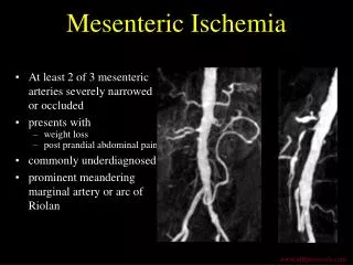
Acute Mesenteric Ischemia:Angiogram Absence of opacification from branches of the aorta (SMA and CA) Images taken from: Uhrad.com-Body Imaging Teaching Files on acute mesenteric ischemia (http://www.uhrad.com/ctarc/ct081.htm)
Surgery • Goals of surgical therapy: • Restore mesenteric blood flow • Assess intestinal viability • Resect nonviable or necrotic intestine