Download
1 / 47
490 likes | 661 Views
Stroke Care Beyond the 3 Hour tPA Window: What are the Options, and How Can They be Accessed?. 2006 Advanced Emergency & Acute Care Medicine and Technology Conference. Emergency Medicine Associates Atlantic City, NJ September 26-27, 2006. Michael A. Ross MD FACEP

E N D
Stroke Care Beyond the 3 Hour tPA Window:What are the Options, and How Can They be Accessed?
2006 Advanced Emergency & Acute Care Medicine and Technology Conference
Emergency Medicine AssociatesAtlantic City, NJSeptember 26-27, 2006
Michael A. Ross MD FACEP Associate Professor Emergency Medicine Department of Emergency Medicine William Beaumont Hospital Wayne State University School of Medicine
Disclosures • All past advisory board or speakers’ bureau activities have expired within the past year
Case Presentation • A 28 year old male presents to the ED as a possible seizure and altered mental status. • His symptoms were first noticed about 3 hours ago. His friends thought he was playing around so delayed transport until he became unresponsive. • He was able to open his eyes, but only make guttural speech. He was not able to sit or stand, and seemed weak on his right side.
Case Presentation • R.O.S. - positive for a mild head ache. • PMHx - essentially negative. • Medications – none • Social - He does not smoke, drink alcohol, or use street drugs. • Family history - non-contributory.
Case Presentation Physical examination showed a well developed male with normal vital signs. • HEENT - He had a weak gag reflex. No trauma. • CHEST / ABD / EXT - Clear breath sounds, regular heart rhythm without murmurs or gallops, normal abdominal and extremity exam. • NEURO - His eyes are open, though he intermittently has disconjugate gaze. His speech was garbled though appropriate. He had profound weakness of his entire right side with complete paralysis of his lower right face, and he could move but not lift his right arm or leg off the stretcher. He has diminished sensation on the right side as well. There are no visual field deficits. His coordination in his left arm and leg is poor, and can not be assessed on the right.
Case Presentation • His initial head CT was normal, as were his ECG and blood-work (CBC, chem-7, drug screen and alcohol). • His respiratory status deteriorated and he was intubated. Chest X-ray showed good tube placement and was otherwise normal. • He was sent for emergency MRA/MRI which showed a basilar artery thrombosis with early infarction of the pons, left midbrain, and right superior cerebellum. He is outside the 3 hour window. What do you do???
Background - Stroke • More than 750,000 new and recurrent strokes occur in the US each year. • Stroke is the THIRD leading cause of death • National cost of stroke = $51 billion annually! • The leading cause of long term disability. • Many consider stroke to be worse than death.
Stroke Etiologies • 40-50% Large vessel occlusions • ICA, MCA, PCA • 25% Small vessel (lacunar) infarcts • 15% Emboli • 15% Primary intracranial hemorrhage
Topics for tonight: • Reperfusion strategies beyond 3 hours • Intra-arterial lytics? • Mechanical embolectomy? • Imaging strategies – The “penumbra conundrum” • What is the issue? • CTA vs MRI?
Systemic lytics: Bad newsTemporal profile of recanalization after iv tPA: Selecting patients for rescue reperfusion techniques ( Ribo, Stroke. 2006;37:1000-1004) • 179 IV tPA patients • Transcranial doppler to assess reperfusion: • Early = 1 hour • Delayed = 2 hours • Late = 6 hours No rec = no recanalize • 39% never reperfuse • Most reperfuse in the first hour (70%) or two (89%)
Systemic thrombolysis:Bad news • Estimated use of IV thrombolysis in lytic eligible U.S. patients : • = 1% - 7% • Reasons for failure to treat: • Leading reason: • Inability to receive and treat within 3 hours • Other reasons: • Fear of IC bleed • CT scan access • Inadequate / uninformed ED personel
Intra-arterial Recanalization • 3-5 hours beyond the traditional 3 hour window. • Best suited for occlusion of a major artery: • The group that tPA does worse in • Major artery mortality is higher: • ICA-T: 53% Jansen, 1995 • MCA: 30-35% Chambers, 1987 • Basilar: 89-92% Bruckman H, 1986 & Brandt, 1996 • Endovascular options: • Intraarterial (IA) tPA • Systemic tPA followed by IA approach • Mechanical revascularization
Intra-arterial thrombolysis(Lisboa, Stroke. 2002 Dec;33(12):2866-71) • Analysis of 27 studies • 852 IA vs 100 control • Outcomes • Good clinical outcome (IA OR = 2.4) • IA = 41% • Control = 23% • Mortality rate • IA = 27% • Control = 40% • Symptomatic ICH • IA = 9.5% • Control = 3%
Intra-arterial thrombolysis(Lisboa, Stroke. 2002 Dec;33(12):2866-71) • Subgroup analysis – favorable outcome: • IA plus IV lytics vs IA (p=0.1, NS.) • IA+IV = 54% • IA alone = 42% • Stroke location (p=0.001) • Supra-tential = 42% • Infra-tentorial = 26%
IMS Study Stroke. 2004;35:904-912 • 80 patients treated with both IV tPA and IA tPA • Historical controls from NINDS trial used for placebo control and IV only. • Outcomes comparable to IV tPA • IC bleed rate same (6.3%) • 3 month “Good outcome” (mRS 0-2) same (43%) • IV then IA lytics is probably safe • Multi-center validation needed = IMS III
Mechanical Revascularization • Techniques • Direct thrombectomy (craniotomy) • Balloon angioplasty • Snare • Embolectomy: MERCI Retriever • Guidewire maceration combined with lytics • Photoacoustic assisted thrombolysis (EPAR) • Ultrasonic intravascular assisted thrombolysis (EKOS)
Mechanical Embolectomy in Acute Ischemic Stroke (MERCI trial) (Smith et al. Stroke, 2005;36:1432-1440)
Multi – MERCI tPA plus MERCI vs MERCI No increased risk when used with IV tPA(July 2006 AJNR)
Now for the big question: • Question: • Beyond 3 hours, how do we know if the infarct is reversible or not??? • Answer: • PENUMBRA IMAGING • BUT WHAT KIND???
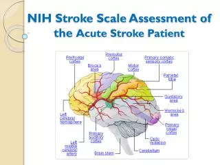
Imaging in acute ischemic stroke – 4 Functions 1. Identify intracranial bleeding • CT is best 2. See if a large or small vessel is occluded • MRA or CTA 3. Identify how much brain is irreversibly damaged • Diffusion MR or CTA 4. Identify clinically relevant penumbra • Perfusion MR or Perfusion CTA
Reperfusion based on penumbra • Viable penumbra was at least as predictive of outcome as time to treatment Ribo M, Molina CA, Rovira A, et al. Safety and efficacy of intravenous tPA stroke treatment in the 3- to 6-hour window using multimodal transcranial Doppler/MRI selection protocol. Stroke 2005;36:602–06
Diffusion weighted MRI • Detects decreased diffusion of water within the brain • Due to altered cell membrane permeability from ischemia. • Hyper-intense signal = non-salvageable tissue • Occurs within minutes of ischemia
Perfusion weighted MRI • Tracks a bolus of gadolinium through the brain. • Cerebral blood flow is indirectly measured • Penumbra is determined by comparing DWI with PWI for mismatch • Traditional approach
MR DWI-PWI conundrum • MR availability • MR technical issues – time, machine, critical patients, metal, etc • MR readings – real time
Comparison of Admission Perfusion CT and Qualitative Diffusion- and Perfusion Weighted MRI in Acute Stroke PatientsWintermark et al. Stroke. 2002;33:2025-2031. Successful Reperfusion Patient Un-successful Reperfusion Patient
Comparison of Admission Perfusion CT and Qualitative Diffusion- and Perfusion Weighted MRI in Acute Stroke PatientsM. Wintermark, MDStroke. 2002;33:2025-2031.
Comprehensive Stroke Centers • Trauma Centers • STEMI hospitals • Comprehensive Stroke Centers??? • STAY TUNED . . .
Case Outcome • The patient was taken to the interventional radiology suite where a MERCI clot retrieval device was successfully used to remove the basilar artery thrombus. • The patient was admitted to the ICU and showed much improvement. • His hospital work-up demonstrated a patent foramen ovale and an underlying hypercoagulable condition. The PFO was patched and the patient was placed on coumadin. ferne_ema_2006_ross_post3hours_092506_final.cd 9/25/2006 5:35 PM
Questions? www.FERNE.org maross@beaumont.edu ferne_ema_2006_ross_post3hours_092606_finalcd 9/25/2006 5:35 PM