Download
1 / 43
3.06k likes | 10.21k Views
NIH Stroke Scale Assessment of the Acute Stroke Patient. NIHSS Assessment of the Acute Stroke Patient . Objectives: The learner will be able to explain Why the NIHSS is performed How the NIHSS is performed Relate each section to the pathophysiology of various types of stroke.

E N D
NIHSS Assessment of the Acute Stroke Patient • Objectives: • The learner will be able to explain • Why the NIHSS is performed • How the NIHSS is performed • Relate each section to the pathophysiology of various types of stroke. • State benefits and drawbacks to the NIHSS • Relate the NIHSS to patient acute changes and long-term outcomes
NIHSS Abstract (Spilker, et. al) “The stroke patient is acutely ill within minutes of symptom onset. Typically, he or she is awake and thus requires a focal neurologic exam to evaluate vision, movement, sensation, and language. With the advent of acute stroke treatments that need to be rapidly implemented, it is critical that the nurse be able to assess patients and relay the information accurately and efficiently to other members of the health team. …. The National Institutes of Health Stroke Scale (NIHSS) is a systematic assessment tool designed to measure the neurologic deficits most often seen with acute stroke patients. “
Case A 43-year old woman is transported by EMS to the ED. Paramedics noted her inability to move the left side of her body. Vital signs on arrival are blood pressure 152/106, temperature 99.2o, heart rate 76, respiratory rate 18. She is awake, oriented to person, place, time and situation. Her husband states she was in another room studying when, at about 8:00pm she called to him complaining of dizziness, a feeling of passing out and left-sided weakness and numbness. The patient is able to provide a partial history of surgery for tetralogy of Falot as a child and recent pacemaker placement (3 years ago). ED staff members made the diagnosis of stroke and implemented the acute stroke protocol. After ED review of all diagnostic data for contraindications. it was determined she was a candidate for thrombolytic therapy.
Arousal Exam versus Focal Neurologic Assessment The patient described above is acutely ill. She has suffered an ischemic stroke in her right middle cerebral artery (MCA) circulation probably secondary to an embolic event from the heart. She is awake and her Glasgow Coma Scale is 15 or normal, her pupils are equal round and reactive to light but despite these ‘normal’ general neurologic findings, she has profoundly abnormal focal neurological signs. However, the GCS and similar tools provide little information about the nature or degree of focal neurological deficits seen in ischemic stroke patients. The neurological exam needed to assess this patient should evaluate the function of this woman’s right cerebral cortex where her neurologic pathology originates.
NIHSS - Background The NIHSS is a systematic assessment tool designed to measure the neurologic deficits more often seen with acute stroke. It was developed in 1983 by NIH-sponsored stroke research neurologists. It was designed to standardize and document an easy to perform, reliable and valid neurologic assessment for use in stroke treatment research trials. Each assessment item was considered for its value during the first hours and days after symptom onset.
General Directions for Performing the NIHSS • Administer scale in numerical order – don’t go back and change scores. • Score what you see, not what you think you should see. Examiners experience may vary and this helps keep assessment objective and consistent. • Minimize cuing or coaching – score first attempt. (Some patients may later correct an error, but do not change the score). • Special considerations – Patients who have difficulty staying awake are more difficult to evaluate due to minimized cooperation. Alternative assessment techniques may be utilized (see further description by item).
Directions for Performing the NIHSS – Aphasic Patients The NIHSS does encourage the use of pantomime and gestures when assessing the aphasic or confused stroke patient. Specific pantomime or gesture clues have been developed with the tool to standardize the cueing that may be necessary to successfully examine and score these patients.
NIHSS Category 1 – Level of Consciousness In acute stroke patients, decreased wakefulness can indicate clinical deterioration. Metabolic causes, due to a generalized or local depression of cerebral cortex cellular function, include hypoxia, systemic organ failure, toxins, infections or ischemia. Structural causes include injury or compression of the brainstem or reticular activating system, either from mass lesions, hematomas, or cerebral edema.
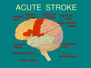
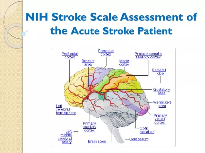
NIHSS Category 2 – Best Gaze “Best Gaze” evaluates selected extraocular movements. Abnormal findings can indicate pathophysiology involving frontal lobe, cerebellar, or vestibular dysfunction, or generalized dysfunction of cerebral cortex.
NIHSS Category 3 – Visual – Visual field assessment Pathologic findings of this item in stroke patients usually arise from lesions of the optic radiations or occipital lobes.
NIHSS Category 4 – Facial Palsy Pathologic findings in item 4 indicate a lower motor neuron lesion of ipsilateral cranial nerve VII or a lesions of the contralateral corticobulbar tract or motor cortex. Bilateral findings can indicate brainstem lesions. This item evaluates symmetry or equality of facial movement. Facial asymmetry can often be seen in even minor stroke. It can be the first clue to the presence of swallowing difficulties or dysphagia.
NIHSS Category 5 – Motor Arm and 6 – Motor Leg Dysfunctions found on items 5 or 6 in stroke patients most often are related to lesions of the motor cortex or corticospinal tract contralateral to the affected side.
NIHSS Category 7 – Limb Ataxia Examines the stroke patient’s ability to coordinate movements. A weakened extremity can appear poorly coordinated but item 7 is scored as present only if the ataxia or poorly controlled movements are out of proportion to the extremity weakness noted in item 5 or 6. Abnormal findings on item 7 often indicate posterior circulation lesions, specifically lesions involving the cerebellum or its connections with the posterior column or brainstem.
NIHSS Category 7 – Limb Ataxia The score is “0” for a paralyzed who cannot move or confused patient who cannot move their extremities or a confused patient who cannot follow directions. This approach increases the tool’s reproducibility,
NIHSS Category 8 – Sensory Sensory deficits and perceptual deficits are common in stroke patients and need to be carefully evaluated to assure patient safety. Abnormal findings or sensory loss usually indicate lesions or dysfunction involving the contralateral thalamus or parietal lobe cortex.
NIHSS Category 9 – Best Language Language deficits are known to be common in stroke patients. Disturbances in speech and communication most commonly indicate lesions in the Broca’s area, Wernicke’s area or the frontal, parietal, or parieto-occipital areas of the left hemisphere. A small number of patients will have language function located in the right hemisphere.
NIHSS Category 9 – Best Language The NIHSS uses a standardized set of visual stimuli.
NIHSS Category 10 – Dysarthria Evaluates quality of the patient’s speech. Pathophysiology is much the same as for facial weakness and can involve either the ipsilateral lower motor neuron cranial nerve deficits or lesions of the contralateral motor cortex. “Dysarthria is a motor speech disorder. The muscles of the mouth, face, and respiratory system may become weak, move slowly, or not move at all after a stroke or other brain injury. The type and severity of dysarthria depend on which area of the nervous system is affected.” http://www.asha.org/public/speech/disorders/dysarthria.htm
NIHSS Category 11 – Extinction and Inattention This item primarily evaluates the contralateral parietal lobe cortex. The ability to perceive needs to be evaluated, documented and considered in the plan of care as early as possible, in ED, ICU or unit. Recognition of visual, tactile, spatial, or personal inattention can prevent falls, one common complications after stroke. Extinction To test extinction, have the patient sit on the edge of the examining table and close their eyes. Touch the patient on the trunk or legs in one place and then tell the patient to open their eyes and point to the location where they noted sensation. Repeat this maneuver a second time, touching the patient in two places on opposite sides of their body, simultaneously. Then ask the patient to point to where they felt sensation. Normally they will point to both areas. If not, extinction is present. With lesions of the sensory cortex in the parietal lobe, the patient may only report feeling one finger touch their body, when in fact they were touched twice on opposite sides of their body, simultaneously. With extinction, the stimulus not felt is on the side opposite of the damaged cortex. The sensation not felt is considered "extinguished".
NIHSS Clinical Performance and Utility • Performing the NIHSS has been timed to take 5 – 8 minutes. • Experience or familiarity increases efficiency. • Patients with impaired attention or language deficits require more exam time even with an experienced examiner. • The complete scale should be done as a baseline and the beginning of each nursing shift. • LOC and extremity motor assessments or specific items designated by the neurologist can be used for more frequent assessments.
NIHSS Clinical Performance and Utility • The NIHSS is used to • Clearly document neurologic outcomes • Plan safe nursing care • Provide consistency of communication between nurses and other health care professionals. • Laminated cards of the visual cues should be readily available • A reference copy of the expanded directions should be stored on every unit that cares for stroke patients. • The general directions should be printed on every version used at the bedside.
Case Study - Revisited It took 6.5 minutes to perform the NIHSS on our patient. • 1. LOCa-0,LOCb-0,LOCc-0 • 2. Best Gaze = 1 • 3. Visual fields = 2 • 4. Facial Palsy = 2 • 5. Motor Arm L=2, R=1 • 6. Motor Leg L=1, R=0 • 7. Limb Ataxia =0 • 8. Sensory =2 • 9. Best Language =0 • 10. Dysarthria = 1 • 11. Extinction/Inattention = 2 TOTAL Score = 14
Meaning of the NIHSS score • The absolute score (0 to 42) has limited meaning. • Think of the score as a way to quantify findings so that changes over time can be measured relative to the individual patient’s baseline. • Findings for sensory, visual, and perception (neglect/ inattention) mean the patient cannot perceive there own deficits or surroundings, thus presents a patient safety risk. • Predictive value for planning rehab or long-term needs • More than 80% of patients with score of 5 or less are discharged home • Those with scores between 6 and 13 usually require acute rehab • Patients with scores of 14 or higher frequently need long-term care. • Patients with low scores may still have a disabling or life-threatening infarction in the cerebellum or brainstem,. • The value of the score does not identify the cause of the stroke. It does not replace a complete neurological exam.
Zero on the NIHSS Does Not Equal the Absence of Stroke • Martin-Schild, et. Al (2011) identified 20 patients with NIHSS = 0 in a population of 2618 patients with acute ischemic stroke admitted to the hospital. • Despite a zero score, these patients had an acute infarction identified by MRI. • The most common presenting symptoms were headache, vertigo, nausea and ataxia. • Neurologic signs on the comprehensive neuro exam included truncal ataxia (45%), agitated confusion (10%), normal exam result (no s/s), and individuals s/s such as nystagmus, limb weakness, memory impairment, Horner's syndrome, reduced visual acuity without field cut. • 57.9% had posterior circulation infarcts, while 42.1% had anterior circulation infarct locations.
What change in NIHSS defines Neurologic Deterioration? Neurologic deterioration (ND) occurs in 1/3 of all acute ischemic stroke (AIS) patients. Recognizing acute changes is crucial in order to address reversible causes of ND Siegler, et. Al (2012) studied 347 patients presenting to one center within 48 hours of onset of AIS. An increase of greater or equal to 2 points in NIHSS was a highly sensitive predictor for poor functional outcome, unfavorable discharge, and mortality. One caveat: an increasing score in a single item (new onset hemiplegia) may call for an emergent workup, even if other improving items (language) offset the change yielding low or zero net effect in NIHSS.
NIHSS, Nursing Care and Patient Outcomes • It is the nurse’s ability to assess the neurologic status of the acute stroke patient and detect changes that will be key to identifying and affecting patient outcomes. • One strategy to build confidence is for the on-coming shift nurse to perform the independent NIHSS and then compare their results to those of the off-going shift. This gives both care providers a chance to compare notes, as well as ensuring a safe handoff. • The nurse should consider the limitations to the NIHSS when identifying a possible stroke patient and in evaluating neurological deteriorization. • Using the NIHSS provides consistent practice throughout the organization and facilitates interdisciplinary coordination.
References Spiller, J., Kongable, G., Barch, C., Braimah, J., Brattina, P., Daley, S., Donmarumma, R., Rapp, K. Sailor, S., and the NINDS rt-PA Stroke Study Group (1997). Using the NIH Stroke Scale to Assess Stroke Patients. Journal of Neuroscience Nursing, 29(6),,394-92. Meyer, B. & Lyden, P. (2009). The Modified National Institutes of Health Stroke Scale (mNIHSS): Its Time Has Come. International Journal of Stroke 4(4). Doi.10.1111/j.1747-4949.2009.00294.x Seigler, J., Boehme, A., Kumar, A., Gillette, M., Albright, K. and Martin-Schild, S. (2012). What Change in the National Institutes of Health Stroke Scale should define neurological deterioration in acute ischemic stroke? Journal of Stroke and Cerebrovascular Disease. Doi:10.1016/j.strokecerebrovasdis.2012.04.012 Martin-Schild, s., Albright, K., Tanksley, J., Panday, V., Jones, E., Grotta, J.m & Savitz, S. (2011). Zero on the NIHSS does not equal the absence of stroke. Annals of Emergency Medicine, 57(1). doi:10.1016/j.annemergmed.2010.o06.564 Kasner, S. (2006). Clinical interpretation and use of stroke scales. Lancet, 5, 603-12.