Download
1 / 42
450 likes | 591 Views
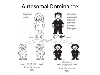
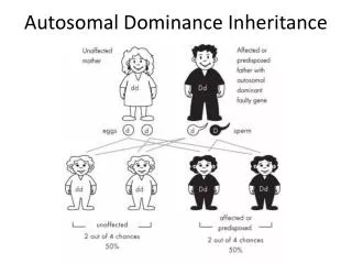
Autosomal Dominant Inheritance. Human Genetics. Autosomal dominant inheritance occurs when one copy of an allele is sufficient for expression of a trait and the gene is located on one of the 22 autosomes. Genetics, Disease, and Dentistry.

E N D
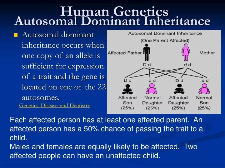
Autosomal Dominant Inheritance Human Genetics • Autosomal dominant inheritance occurs when one copy of an allele is sufficient for expression of a trait and the gene is located on one of the 22 autosomes. Genetics, Disease, and Dentistry Each affected person has at least one affected parent. An affected person has a 50% chance of passing the trait to a child. Males and females are equally likely to be affected. Two affected people can have an unaffected child.
Autosomal Dominant Disorders • Progeria: characterized by an appearance of accelerated aging in children, affects 1 in 8 million newborns • Huntington’s Disease: degenerative brain disorder, slowly diminishes the affected individual’s ability to walk, think, talk and reason dementia
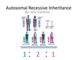
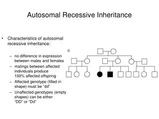
Autosomal Recessive Inheritance • Autosomal recessive inheritance occurs when two copies of an altered gene located on one of the autosomes must be present for an individual to be affected with the trait or condition determined by that gene. • An affected individual (homozygote) has two parents who are unaffected but each parent carries the altered gene (heterozygote). • Males and females are at equal risk for being affected. • Two affected individuals usually produce children all of whom are affected as well.
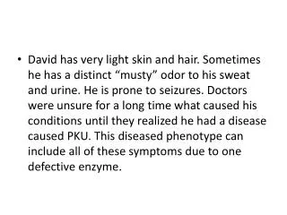
Autosomal Recessive Disorders • Tay-Sachs Disease: individuals lack an enzyme in the lysosomes of their brain cells needed to break down lipids. The undigested lipids enlarge and eventually destroy the brain cells that house them. • Phenylketonuria (PKU): individuals lack an enzyme that converts Phe to Tyr. Failure of the conversion to take place results in a buildup of Phe. Through a mechanism that is not well understood, the excess Phe is toxic to the central nervous system. This results in mental retardation and other neurological problems if not detected early. • Albinism Sickle Cell Anemia
X-Linked Inheritance • Most individuals who are affected with the trait or condition in questions are males. • Mothers of the affected males are carriers and the sisters of affected males may be either carriers or not carry the gene al all. The basis for X-linked inheritance is that females have two X chromosomes and males have only one X chromosome. • Affected males are hemizygous (their one X chromosome has the mutant allele) • Affected females are homozygous (both X chromosomes have the mutant allele) • Affected males transmit the gene to all daughters, but not to any of their sons • The daughters of an affected male will usually be a carrier (heterozygote) and thus not show the trait (masked) • Sons of heterozygous females have a 50% chance of receiving the gene and thus expressing the trait or condition
X-linked Disorders • Colour Blindness • In red-green colour vision deficiency, the visible spectrum is divided into two parts; a red segment and a blue segment, separated by grey or indistinct areas. The amount of grey or indistinct areas varies according to the severity of the deficiency. • Men are mainly affected • For a woman to be colour deficient, her father must be colour-blind and her mother must be a carrier • A defective male always inherits his deficiency from his mother who usually has normal colour vision is therefore a carrier of the defect.
Duchenne Muscular Dystrophy • Involves the wasting away of muscle tissue • Muscle cells become engorged with fat and they eventually waste away most individuals suffer from respiratory failure in their early twenties.
Human Pedigrees: Working out Inheritance Patterns Factors to Consider in Pedigrees • Is the trait located on a sex chromosome or an autosome? • Autosomal – not on a sex chromosome • Sex Linkage – located on one of the sex chromosomes • Y-linked - only males carry the trait. • X-linked (recessive) - sons inherit the disease from normal parents • How is the trait expressed? • Dominant - the trait is expressed in every generation. • Recessive - expression of the trait may skip generations. Pedigrees are a convention for keeping track of human genetic traits used to infer genotype. Pedigrees are the human equivalent of test crosses. In a visualization of a pedigree: males are designated with square symbols. females with round symbols lines are drawn to indicated matings, parent-offspring relationships, and relationships between siblings.
Pedigree Diagrams: I Pedigree Diagrams: II • Basic Symbols • Basic Symbols for offspring and the expression of a trait. • The offspring are depicted below the parents. • Filling the symbol with black indicates the expression of the studied trait. Pedigree analysis
Note that the symbols for non-identical twins and for identical twins differ by whether they descend from a common vertical before bifurcating Media Showcase
Pedigrees • Generations are numbered from the top of the pedigree in uppercase Roman numerals, I, II, III etc. Individuals in each generation are numbered from the left in arab numberals as subscripts, III1 , III2, III3 etc. For example, here is a typical autosomal dominant pedigree with numbered generations and individuals.
Marfan’s Syndrome: An Example • Expressed in both sexes. • Thus, autosomal. • Expressed in every generation. • Thus, dominant.
Marfan’s: Genotype the Normal Individuals • Assign codes for the alleles. • Code “m” for the recessive normal allele. • Code “M” for the dominant allele for Marfan’s syndrome. • Normal individuals must be “mm.”
Marfan’s: Genotype the Affected Individuals • Affected individuals must have at least one “M.”
Marfan’s: Parent-Offspring Relationships • Possibilities for #1 and #2: Heterozygote (Mm) or homozygous for “M?” • If “MM,” all offspring from a normal mate should be affected. • Therefore, both must be heterozygotes.
Marfan’s: Parental Genotypes Known • “M” must have come from the mother. • The father can contribute only “m.” • Thus, the remaining genotypes are “Mm.”
Albinism: An Example • Expressed in both sexes at approximately equal frequency. • Thus, autosomal. • Not expressed in every generation. • Thus, recessive.
Albinism: Genotype the Affected Individuals • Assign codes for the alleles. • Code “A” for the dominant normal allele. • Code “a” for the recessive allele for albinism. • Affected individuals must be homozygous for “a.” • First generation parents must be “Aa” because they have normal phenotypes, but affected offspring.
Albinism: Genotype the Normal Individuals • Normal individuals must have at least one “A.”
Albinism: Parent-Offspring Relationships • #1 must transmit “a” to each offspring. • The “A” in the offspring must come from the father. • Normal father could be either heterozygous or homozygous for an “A.”
Albinism: Parental Genotypes are Known • Both parents are heterozygous. • Normal offspring could have received an “A” from either parent, or from both.
Albinism: One Parental Genotype is Known • Only the genotype of the offspring expressing albinism are known. • Normal offspring must have received an “a” from their affected father.
Hairy Ears: An Example • Only males are affected. • All sons of an affected father have hairy ears. • Thus, hairy ears is Y-linked.
Hairy Ears: Female Sex Determination • All females are XX
Hairy Ears: Male Sex Determination • All males are XY.
Hairy Ears: Gene on the Y Chromosome • Code “H” indicates the allele on the Y chromosome for hairy ears
Hairy Ears: Wild-Type Allele for Normal Ears • Code “+” indicates the allele on the Y chromosome for normal ears.
Hemophilia: An Example • In this pedigree, only males are affected, and sons do not share the phenotypes of their fathers. • Thus, hemophilia is linked to a sex chromosome–the X. • Expression of hemophilia skips generations. • Thus, it is recessive. Children resemble their parents.: Animation Extensive bruising of the left forearm and hand in a patient with hemophilia.
Hemophilia: Expression of the Female Sex Chromosomes • All females are XX
Hemophilia: Expression of Male Sex Chromosomes • All males are XY.
Hemophilia: Genotype the Affected Individuals • Assign codes for the alleles. • Code “H” for the recessive hemophilia allele. • Code “+” for the wild-type normal allele. • Affected individuals must have an “H” on an X chromosome.
Hemophilia: Father-Daughter Relationship • All daughters of an affected father receive an X chromosome with the “H” allele.
Hemophilia: Genotyping the Normal Individuals • Normal individuals must have at least one X chromosome with the wild-type allele, “+.”
Hemophilia: Homozygous or Heterozygous? • Only males affected • Not Y-linked • Skips a generation: recessive • X-linked
Try It! • Let us begin by drawing the pedigree described below (which is not necessarily an autosomal dominant condition and which contains extraneous information).
The Scenario • Alice and Bob have a two year old son, Charles, who is showing mental retardation, short stature, micropenis, and cryptorchidism. Alice has two living, unaffected, brothers but her eldest brother died at age 9 and a second brother died aged 10 months. Both had similar problems to Charles. Alice's father, David, who was symptomless, has a sister, Ethel, who has an unaffected boy and girl, and a brother, Fred, who also has two unaffected children. Alice's mother, Gertrude, has two living sisters and had a brother who had died in childhood and who, she remembers, had been mentally retarded. Bob has two brothers, Henry and Ignatius, who are still unmarried. His parents, John and Kate, had tragic lives, both were adopted and never knew their biological parents and both died as the result of a road accident.
Step 1 • Begin with Alice, Bob and Charles. • Here are three possible drawings of this nuclear family.
Correct Solution • Alice and Bob are connected by a horizontal line to show that this is a mating. Charles is connected to that horizontal line to show that he is a product of that mating.
Now add Alice's siblings and parents to the pedigree. Choose One… Step 2
Correct Solution was Possiblilty 3 Alice and her four brothers are connected vertically to a horizontal line which is, in turn, connected to the line drawn between her parents David and Gertrude. Her two dead brothers (whom we presume died of the same genetic disease - though this can sometimes be a foolish assumption without medical evidence) are shaded in (to show that they suffered from the disease) and are crossed through (to show that they are dead).
Step 3 • Now add Gertrude's siblings to the pedigree. • And David's siblings and his nephews and nieces • Finally add Bob's side of the family • Try Drawing it!!!
Correct Drawing! http://bio1151.nicerweb.net/med/Vid/Discover2e/ch13a04_Pedigree.swf Children resemble their parents.: Problem