Download
1 / 66
660 likes | 702 Views
THE MUSCULAR SYSTEM: SKELETAL MUSCLE TISSUE AND MUSCLE ORGANIZATION. Muscle Tissue: Functions . 1. producing body movements integrated action of skeletal muscle, joints and bones 2. stabilizing body positions skeletal muscle contraction stabilizes joints and bones

E N D
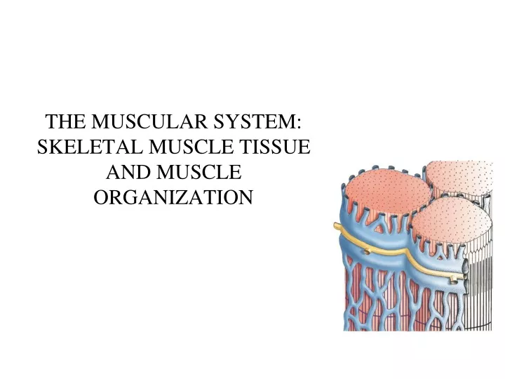
THE MUSCULAR SYSTEM: SKELETAL MUSCLE TISSUE AND MUSCLE ORGANIZATION
Muscle Tissue: Functions • 1. producing body movements • integrated action of skeletal muscle, joints and bones • 2. stabilizing body positions • skeletal muscle contraction stabilizes joints and bones • postural muscles contract continuously when awake • 3. maintain body temperature • 4. storing and moving substances in the body • contraction of ring-like smooth muscle sphincters – storage of material in an organ • regulation of entrance and exit to and from the body • storage of glucose within skeletal muscle • movement of blood by cardiac muscle and by smooth muscle within the blood vessels • movement of food through the GI tract by smooth muscles within abdominal viscera
Properties of Muscles Electrical excitability -ability to respond to stimuli by producing electrical signals known action potentials -requires the receipt of a stimulus by the muscle cell -two types of stimuli: 1. electrical signals 2. chemical stimuli = neurotransmitters Contractility -ability to contract when stimulated by an AP -isometric contraction: tension develops, length doesn’t change -isotonic contraction: tension develops, muscle shortens Extensibility -ability to stretch without being damaged -allows contraction even when stretched Elasticity -ability to return to its original length and shape
Gross Anatomy • muscles are really groups of fascicles • the fascicles are groups of muscle fibers • muscle fiber is considered to be an individual muscle cell • muscles are covered superficially by a superficial fascia layer (or subcutaneous layer/hypodermis) • three layers of connective tissue surround the muscle & partition it into bundles and fibers • Epimysium • Perimysium • Endomysium • these layers further strengthen and protect muscle • outermost layer = epimysium • encircles the entire muscle • next layer = perimysium • divides the muscle into bundles of 10 to 100 individual muscle fibers • fibers = muscle cell • give meat its “grain” because the fascicles are visible • both epimysium and perimysium are dense irregular connective tissue
next layer = endomysium (areolar connective tissue) • penetrates the fasicles and separates them into individual muscle fibers • skeletal muscle fibers are controlled by lower motor/somatic motor neurons = neuromuscular junction • the muscle fiber is made up of fused muscle cells -these muscle cells have a unique cytoskeleton made up of myofibrils
each myofibril is comprised of repeating units of protein filaments = sarcomeres
Skeletal Muscle development • Large, multinucleated cells • embryonic development - muscle fibers arise from fusion of a hundred or more mesodermal cells called myoblasts – organized into muscle fibers • once fused, these muscle cells lose the ability of undergo mitosis • number of muscle cells predetermined before birth • myoblasts develop from stem cells • adult – these stem cells are called satellite cells • also can come from bone marrow stem cells? muscle fibers
The muscle cell: contents • Plasma membrane = sarcolemma • nuclei • organelles • golgi, lyososome • mitochondria • sarcoplasmic reticulum • T-tubules • Cytoplasm = sarcoplasm • cytoskeleton • microtubules • intermediate filaments (desmin) • actin • myosin • other structural proteins • cytosol • water • ions • glycogen (glucose) • myoglobin (oxygen) • creatine phosphate (ATP)
Microanatomy of Muscle Fibers • New terminology • Cell membrane = sarcolemma • plasma membrane that surrounds a single muscle fiber • Cytoplasm = sarcoplasm • substantial amounts of glycogen - can be broken into glucose • contains myoglobin - binds oxygen needed for muscle ATP production
Internal membrane system = sarcoplasmic reticulum • encircles each myofibril • have dilated end sacs = terminal cisternae • stores calcium when at rest • releases it during contraction • calcium release is triggered by an AP • Invaginations of the plasma membrane = T tubules • open to the outside of the fiber and continuous with the sarcolemma • filled with interstitial fluid • action potentials travel along the sarcolemma and the T tubules • allows for the even and quick spread of an action potential Microanatomy of Muscle Fibers
M line Sarcomere Structure • sarcomere = regions of myosin (thick myofilament) and actin (thin myofilament) • bounded by the Z line • about 1.6 to 2.0 um in length • typical myofibril has about 10,000 sarcomeres • comprised of an A band, an H zone, and 2 halves of an I band
Sarcomere structure • I band – region of thin filaments only • split in half by the Z line • Z line – denotes the sarcomere • comprised of proteins called connectins (e.g. actinin) that interconnect the thin filaments from sarcomere to sarcomere • also connect to a structural protein called titin • keeps thick and thin filaments in proper alignment and resists extreme stretching forces • connected to adjacent myofibrils by intermediate filaments (e.g. desmin) • results in the sarcomeres from adjacent myofibrils aligning with each other = striated appearance • M line – made up of a protein called the M line protein • binds an enzyme for ATP storage called creatine kinase • helps in the positioning of the thick filaments between the thin filaments (binds myosin) • A band – length of the thick filament • contains the “zone of overlap” between thin and thick filaments
The Proteins of Muscle • Myofibrils are built of 3 kinds of protein • contractile proteins • myosin and actin • regulatory proteins which turn contraction on & off • troponin and tropomyosin • structural proteins which provide proper alignment, elasticity and extensibility • E.g. titin, nebulin and dystrophin
Structural Proteins of Muscle • Nebulin, an inelastic protein helps align the thin filaments. • Dystrophin links thin filaments to sarcolemma and transmits the tension generated to the tendon. • Titin anchors thick filament to the M line and the Z line.
Muscular dystrophy • genetic disorder of muscle weakness and defects in muscle proteins • several types recognized • Duchenne’s: childhood onset • X linked • less severe effects in females • dystrophin – helps anchor cytoskeleton to PM • mutant dystrophin incapable of properly transmitting contractile force of the sarcomere to the plasma membrane = muscle weakness
Contractile proteins of muscle: Actin • two forms of actin – G-actin and F-actin • the G-actin “beads” are assembled together (using ATP) to form two linear chains of actin called F-actin • these two chains are wrapped around a core “rod” of nebulin to form a helix • F-actin filament is associated with the regulatory proteins troponin and tropomyosin = THIN FILAMENT
Contractile proteins of muscle: Myosin • myosin thick myofilament is a bundle of myosin molecules • each myosin protein is made up of two heavy chains - each with a globular “head” with a site to bind ATP and a site to bind actin • also associated with the heads are 4 light chains • play a role in myosin’s assembly and ability to hydrolyze ATP
Contraction: The Sliding Filament Theory • Contraction: • Active process • Elongation is passive • Amount of tension produced is proportional to degree of overlap of thick and thin filaments • SF Theory: • Explains how a muscle fiber exerts tension • Four step process • Active sites on actin • Crossbridge formation • Cycle of attach, pivot, detach, return • Troponin and tropomyosin control contraction
Calcium binds troponin tropomyosin Recovery ATP hydrolysis Power stroke -calcium binds to troponin and exposes sites that can interact with myosin - Ca+2 binds to troponin & causes troponin-tropomyosin complex to move & reveal myosin binding sites on actin -ATP binding, hydrolysis and ADP release changes the conformation of the head (“power stroke”) and causes actin to “slide” along the myosin myofilaments -shortens the distance between the Z lines
Myosin head = ADP Myosin head = empty Myosin head = ATP Myosin head = ADP CHECK OUT THIS ANIMATION!!! http://www.blackwellpublishing.com/matthews/myosin.html
CHECK OUT THESE ANIMATIONs!!! https://www.youtube.com/watch?v=Ktv-CaOt6UQ http://highered.mcgraw-hill.com/sites/0072495855/student_view0/chapter10/animation__myofilament_contraction.html http://www.youtube.com/watch?v=EdHzKYDxrKc http://www.youtube.com/watch?v=Vlchs4omFDM http://www.youtube.com/watch?v=Ct8AbZn_A8A http://www.youtube.com/watch?v=BMT4PtXRCVA https://www.youtube.com/watch?v=4201SrN0WlY
Sarcoplasmic Reticulum and Calcium release • the SR wraps around each A and I band • segmented with T-tubules between each SR segment • each segment forms saclike regions at the ends = lateral sacs (terminal cisternae) • site of calcium storage
T-Tubules and the Sarcoplasmic Reticulum • The T-tubule system of muscle cells is for the conduction of the action potential deep inside the cardiomyocyte • The T-tubule is flanked on both sides by the sarcoplasmic reticulum • The passage of the action potential through a T-tubule causes the release of calcium from the sarcoplasmic reticulum (lateral sacs) T-tubule Sarcoplasmic reticulum
The AP and contraction • “slow” L-type Ca+ channels are found within the T-tubules • also known as dihydropyridine receptors • opening these triggers the opening of Ca+ channels within the adjacent lateral sacs of the sarcoplasmic reticulum • these calcium channels are known as ryanodine or foot proteins (type RyR1) • opening results from the physical interaction between the dihydropyridine receptors with their ryanodine “partners” • this triggers a very large release of Ca+ from the SR = “Ca-induced Ca release” • as more dihydropyridine receptors interact with more ryanodine receptors • burst of Ca+ = Ca+ sparks • together with the slow removal of Ca+ - results in a long sustained contraction of heart muscle
The Neuromuscular Junction • end of neuron (synaptic terminal or axon bulb) is in very close association with a single muscle fiber (cell) • nerve impulse leads to release of neurotransmitter(acetylcholine) from the synaptic end terminal
The Neuromuscular Junction • AcH binds to acetylchloline receptors on myofibril surface - ligand-gated Na+ channels (ionotropic nicotinic receptors) • binding leads to influx of sodium ions and depolarization of the membrane potential of the sarcolemma • creation of an action potential that travels through the muscle cell – eventual contraction • Acetylcholinesterase breaks down ACh • Limits duration of contraction
Muscle Contraction: A summary • ACh released from synaptic vesicles at each neuromuscular junction • Binding of ACh to muscle cell of the NMJ • entrance of Na+ ions and depolarization • Generation of action potential in sarcolemma • Conduction of impulse along T-tubules • AP flows along the outside of the muscle cell via the sarcolemma • enters the inside of the muscle cell via T-tubules • close association of T-tubules with the sarcoplasmic reticulum (SR) • Release of Calcium ions by SR • AP results in release of Ca2+ by the SR • SR is in close physical association with each A and I band • Exposure of active sites on actin • Ca2+ binds to troponin and “pulls it away” from the actin filament • Cross-bridge formation with myosin • requires hydrolysis of ATP ADP • Release of ADP • pivoting of myosin head • sliding filaments & contraction • also called excitation-contraction coupling – describes the events linking generation of an AP (excitation) to the contraction of the muscle
The Events in Muscle Contraction CHECK OUT THIS ANIMATION!!! http://www.blackwellpublishing.com/matthews/myosin.html
Length of Muscle Fibers: Length Tension relationship • Normally • resting muscle length remains between 70 to 130% of the optimum • Optimal overlap of thick & thin filaments • produces greatest number of crossbridges and the greatest amount of tension • optimal length = lo (muscle length at which maximum force is generated) • optimal length = point A A B D C
Length of Muscle Fibers: Length Tension relationship • As stretch muscle (past optimal length) • length of the muscle fiber is greater than lo • fewer cross bridges exist & less force is produced = point B • when muscle is stretched to about 70% than lo of its (point C) the actin filaments are completely pulled out from between the myosin – no cross-bridges possible • If muscle is overly shortened (less than optimal) • length of the muscle fiber is less than lo • thick filaments crumpled by Z discs and the actin filaments overlap – poor cross-bridge formation • fewer cross bridges exist & less force is produced = point D • even less calcium released from the SR - ?? A B D C
Motor Units • Each skeletal fiber has only ONE NMJ • MU = Somatic neuron + all the skeletal muscle fibers it innervates • Number and size indicate precision of muscle control • Muscle twitch • Single momentary contraction in one muscle fiber • too small to generate any significant force • Response to a single stimulus • All-or-none theory • Either contracts completely or not at all • Motor units are grouped together to provide a greater force & delay fatigue • in a whole muscle they fire asynchronously • some fibers are active others are relaxed • delays muscle fatigue so contraction can be sustained • Muscle fibers of different motor units are intermingled so that net distribution of force applied to the tendon remains constant even when individual muscle groups cycle between contraction and relaxation.
Neural control of Motor Units • 1. input from the motor cortex • axons originating from neuronal cell bodies within the primary motor cortex descend directly to synapse with motor neurons in the SC • part of the corticospinal motor system (lecture 8) • 2. input from the brain stem • extrapyramidal motor system • involves many regions of the brain • final link is the brain stem
Neural control of Motor Units • 3. input from afferent neurons • at the level of the SC • by interneurons within the SC = spinal reflex • afferent information is needed to control skeletal muscle activity • the CNS must know the position of your body prior to initiating movement and must know how the movement is progressing = prioprioceptive input • comes from information from your eyes, joints, inner ear and from the muscles themselves (prioprioceptors) • muscle spindles and tendon organs within the muscle monitor changes in muscle length and tension (see lecture 9)
Motor Tone • Resting muscle contracts random motor units • Constant tension created on tendon • Resting tension – muscle tone • Stabilizes bones and joints • controlled by proprioceptors
Muscle Metabolism • Production of ATP: -contraction requires huge amounts of ATP -muscle fibers produce ATP three ways: 1. Creatine phosphate 2. Aerobic metabolism 3. Anaerobic metabolism
Creatine Phosphate • Muscle fibers at rest produce more ATP then they need for resting metabolism • Excess ATP within resting muscle used to form creatine phosphate or phosphocreatine • Creatine phosphate: 3-6 times more plentiful than ATP within muscle • the first storehouse of energy used upon the onset of contraction when additional ATP is needed • Sustains maximal contraction for 15 sec (used for 100 meter dash) • or about 8 muscle twitches • creatine phosphate breakdown is favored by muscles undergoing explosive movements • formed through the combination of creatine and ATP • when energy is required – CP splits up into creatinine and a high energy phosphate group • the phosphate group is added to ADP to recreate ATP • creatine phosphate and reconversion both catalyzed by creatine kinase • CK is linked to the M line in sarcomeres
Anaerobic Cellular Respiration • Muscles deplete creatine – make ATP in anaerobically via glycolysis • glucose 2 pyruvic acid • if enough O2 is available to the muscle pyruvic acid made during glycolysis enters the citric acid cycle and electron transport chain to make ATP • BUT: if insufficient oxygen is present only glycolysis is performed • glucose is broken down into two pyruvic acid molecules to yield 2 ATP • BUT in low oxygen this pyruvic acid is further processed to the by-product = lactic acid • Glycolysis can continue anaerobically to provide ATP for 30 to 40 seconds of maximal activity (200 meter race) http://www.indstate.edu/thcme/mwking/oxidative-phosphorylation.html
Aerobic Cellular Respiration • aerobic respiration produces ATP for any activity lasting over 30 seconds • if sufficient oxygen is available, pyruvic acid enters the mitochondria to generate ATP, water and heat via the electron transport chain • fatty acids and amino acids can also be used by the mitochondria • can provide 90% of ATP energy if activity lasts more than 10 minutes is intensity level is moderate • oxygen levels must be sufficient!!!!! • Each glucose = 36 ATP • Fatty acids = ~100 ATP • Sources of oxygen – diffusion from blood, released by myoglobin (hemoglobin-like molecule of muscle cells)
Muscle Fatigue • Inability to contract after prolonged activity • two types: central fatigue and peripheral fatigue • peripheral fatigue – events that happen anywhere between the NMJ and the sarcomere • central fatigue is feeling of tiredness and a desire to stop (protective mechanism) – also called psychological fatigue • Factors that contribute to muscle fatigue = Peripheral Fatigue mechanisms • depletion of creatine phosphate • decline of Ca+2 within the sarcoplasm • insufficient glycogen – may actually affect Ca++ release from the SR • accumulation of extracellular K ions • buildup of lactic acid • may inhibit key enzymes in the coupling of AP to contraction??? role??? • drop in pH within the muscle cell • build up of lactic acid or through ATP hydrolysis • may only occur in extreme exertion – since pH is controlled well in most cells • buildup of ADP and inorganic phosphate from ATP hydrolysis • directly interfere with cross-bridge formation and/or block calcium release and re-uptake by sarcoplasmic reticulum • insufficient release of acetylcholine from motor neurons – disease states only
Isotonic and Isometric Contraction • Isotonic contractions = a load is moved • concentric contraction = a muscle shortens to produce force and movement • eccentric contractions = a muscle lengthens while maintaining force and movement • Isometric contraction = no movement occurs • tension is generated without muscle shortening • maintaining posture & supports objects in a fixed position
Atrophy • wasting away of muscles • caused by disuse (disuse atrophy) or severing of the nerve supply (denervation atrophy) • the transition to connective tissue can not be reversed • Hypertrophy • increase in the diameter of muscle fibers • resulting from very forceful, repetitive muscular activity and an increase in myofibrils, SR & mitochondria
Exercise-Induced Muscle Damage • Intense exercise can cause muscle damage • electron micrographs reveal torn sarcolemmas, damaged myofibrils an disrupted Z discs • increased blood levels of myoglobin & creatine phosphate found only inside muscle cells • Delayed onset muscle soreness • 12 to 48 Hours after strenuous exercise • stiffness, tenderness and swelling due to microscopic cell damage
Three Types of Muscle Fibers • Fast fibers = fast twitch glycolytic • Slow fibers = slow twitch oxidative • Intermediate fibers = fast twitch oxidative-glycolytic • Fibers of one motor unit all the same type • Percentage of fast versus slow fibers is genetically determined • Proportions vary with the usual action of the muscle - neck, back and leg muscles have a higher proportion of postural, slow oxidative fibers - shoulder and arm muscles have a higher proportion of fast glycolytic fibers
Fast Fibers • Large in diameter • Contain densely packed myofibrils • Large glycogen reserves • pump calcium into their SRs faster – faster twitches (single cell contraction rates) • high ATPase activity – faster cross-bridge/contraction potential • fatigue very quickly
Fast Fibers • Fast twitch oxidative-glycolytic (fast-twitch A) • red in color (lots of mitochondria, myoglobin & blood vessels) • higher ability to produce ATP via aerobic metabolism • highly vascularized • split ATP at very fast rate; used for walking and sprinting • lower resistance to fatigue • Fast twitch glycolytic (fast-twitch B) • white in color (few mitochondria & BV, low myoglobin) • higher concentration of enzymes for glycolysis • need less oxygen to function • anaerobic movements for short duration; used for weight-lifting • fatigue faster also
Slow fibers • Half the diameter of fast fibers • Three times longer to contract • low ATPase activity, low glycogen content • high resistance to fatigue • higher ability to produce ATP via aerobic metabolism • many mitochondria • highly vascularized • continue to contract for long periods of time • e.g. marathon runners