Download
1 / 63
630 likes | 660 Views
Sleep and Wakefulness. Jonathan W. Bekenstein, M.D., Ph.D. Associate Professor of Neurology May 21, 2009. OBJECTIVES:. Understand the normal human sleep cycle. Grasp the mechanisms of the circadian clock. Understand the retinohypothalamic pathway.

E N D
Sleep and Wakefulness Jonathan W. Bekenstein, M.D., Ph.D. Associate Professor of Neurology May 21, 2009
OBJECTIVES: • Understand the normal human sleep cycle. • Grasp the mechanisms of the circadian clock. • Understand the retinohypothalamic pathway. • Know EEG characteristics of stages of sleep. • Know the neurotransmitter systems and nuclei in the reticular activating system responsible for transitions to different sleep stages.
Basic Principles • Humans spend about one-third of their lives sleeping. We do not know the entire function of sleep, but many functions are understood. These include energy conservation and restoration of brain glycogen levels. • Metabolism is diminished at night, and so is body temperature, both of which follow a circadian rhythm and generally follow a pattern of ambient temperature.
Developmental Changes • Sleep evolves and changes during life and development. Total sleep requirements change during life. Adolescents and young adults often require more sleep, up to 10 hours per day. Early school schedules often cause adolescents to have daytime sleepiness due to insufficient total sleep time.
Why do we sleep? • Many experts hypothesize that since humans are highly visual beings, lack of visual stimulation and an inability to fully function in the dark may be part of why we sleep. • Sleep may be a way to shed accumulated body temperature from daytime activities and to reduce the production of body heat due to reduced activity. Growth occurs primarily during slow wave sleep in the early night, when pulsatile release of growth hormone is at its highest level. • Temperature regulation-Growth- Memory
Sleep and Immune System • Interleukin-1 disrupts sleep-wake cycle through receptors in brainstem. • May need sleep to help fight infections. • Maintain fever during sleep and alter thermoregulation during infections. • Sleep deprivation impairs leukocyte function and DNA synthesis for up to 5 days.
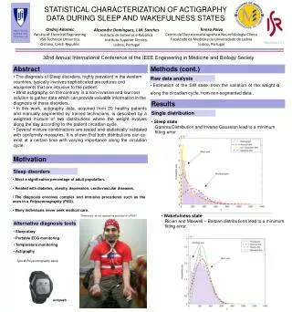
Proposed principles by which changes in sleep architecture promote recovery from infection.
Possible Functions of Sleep • Protein synthesis and RNA transcription are most abundant during slow wave sleep, which may have implications for maintaining synaptic function or making memory more persistent. Lastly, mood and cognition, particularly concentration are adversely affected by sleep deprivation or sleep with frequent interruptions.
Circadian Rhythms and Sleep • (Circa=about; dia-day) Circadian rhythms are those that occur with about a 24 hour periodicity. Humans have an internal “clock” that runs in the absence of external clues about darkness and light. The internal clock “free-runs” with a period of about 24.2 hours, just slightly longer than a day on Earth. The internal clock can quickly be entrained with light shining into the eyes at a set time each day. Special cells in the retina contain a pigment called melanopsin. Axons from these retinal ganglion cells project to the suprachiasmatic nucleus of the anterior hypothalamus.
Paraventricular nucleus Supraoptic nucleus
Phototransduction • Phototransduction from the retina to the suprachiasmatic nuclei occurs primarily via the retinohypothalamic tract. Across a normal waking day, clock-dependent (circadian) alertness is usually lowest in the early morning and increases into the late afternoon or evening, thus opposing the growing sleepiness from having been awake all day.
Pathway for signaling Circadian light changes • Melanopsin containing retinal ganglion cells project to the suprachiasmatic nucleus of the anterior hypothalamus. • SCN projects to the paraventricular nucleus of the hypothalamus. Those cells send axons to the preganglionic sympathetic neurons in the intermediolateral zone in the thoracic spinal cord. • Intermediolateral cells project to the superior cervical ganglion.
Pathways (continued) • Postganglionic axons project to the pineal gland (pinecone shaped). • Melatonin is synthesized from L-tryptophan. • Melatonin levels rise in CSF about two hours before sleep onset and about 7 hours before the core body temperature falls to its nadir. Thus, sleep onset is 5 hours or so before the lowest core body temperature is reached.
What Role Does Light Play? • Light inhibits melatonin production during the day. • Increasing Melatonin is required for sleep. • Serotonin is converted to Melatonin in the Pineal gland.
Synchronization of Circadian Timekeeping among SCN Neurons Pacemaking neurons generate near-24-hr rhythms in gene expression, firing rate, and peptide release through a transcription-translation negative-feedback loop. These neurons are all GABAergic, and a subset in the ventral (core) SCN release VIP. VIP and its receptor VPAC2 are necessary for synchronization of circadian periods among SCN neurons. Daily GABA application can synchronize SCN neurons, and blockade of GABAA receptors interferes with rhythm coordination between the dorsal (shell) and ventral SCN. Gap junctions have also been implicated in spike-for-spike synchrony between neighboring SCN neurons.
Explanation • Robust rhythm generation in SCN neurons is thought to rely on circadian expression of Period (Per1, 2), Cryptochrome (Cry1, 2), Rev-Erb, Ror, and Bmal1 genes, and constitutive expression of Clock. Period and Cryptochrome proteins (PER/CRY) form a negative-feedback loop, repressing transcriptional activation by CLK/BMAL1 through E-Box sequences on Per and Cry promoters. Casein kinase 1 and (CK1Δ) causes a phosphorylation-dependent delay in PER/CRY feedback. REV-ERB and ROR proteins form a second feedback loop, binding to ROR-element (RORE) promoters of the Bmal1 gene to repress and enhance expression, respectively.
More than half of all SCN neurons require daily VIP-VPAC2 signaling to maintain robust rhythms. This signaling pathway may impinge on the intracellular molecular clockwork through activation of adenyl cyclase (AC), cAMP, protein kinase A (PKA), and CREB-dependent transcription of the Period genes (Travnickova-Bendova et al., 2002 and Itri and Colwell, 2003). This regulation of clock gene transcription may underlie the synchronization and amplification of neuronal rhythms by daily VIP/VPAC2 signaling. (Aton and Herzog, Neuron, 2005:531-534)
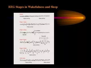
Stages of Sleepand Some Basic EEG • Stages of Sleep • 1. Defined by EEG criteria, as behavioral distinction is difficult. • a. Alpha frequency: 8-13 Hz (described first) • b. Beta: > 13 Hz (described second) • c. Theta: 4-7 Hz • d. Delta < 4 Hz (for Death, Disease, Disability)
There is a progression through stages of sleep. • Drowsiness characterized by diminished eye blinks, diminution and cessation of the posterior dominant alpha rhythm, and some diffuse slowing of the background frequencies. Alpha is the predominant awake rhythm. Beta is seen in the frontal head regions during wakefulness. Theta is a normal rhythm of limbic structures and hippocampus in wakefulness.
More Definitions • Other than in disease states and in damaged brain, Delta waves are associated with non-REM (rapid eye movement) sleep. Sleep spindles are waves of waxing and waning activity that have a fronto-central predominance at a frequency between 7-16 Hz. They are generated by reticular thalamic cells that are GABAergic.
Non-REM Sleep: Stage I: more theta activity, loss of alpha, and vertex sharp waves Stage II: theta and delta waves, vertex sharp waves, K complexes (a vertex sharp wave with a superimposed slow and fast component), and sleep spindles.
Sleep Stages • Stage III: higher voltage delta waves that are 20-50% of the background over time. • Stage IV: more than 50% delta slow waves. • During non-REM sleep muscle tone is maintained at a moderate level and breathing is regular.
REM Sleep • Return of beta activity to the EEG • Loss of muscle tone except in diaphragmatic muscles • Rapid eye movements • Dreaming • Lasts about 10 minutes per epoch, on average. • Total REM time diminishes from 8 hours at birth to 2 hours at age 20 to 45 minutes at age 70.
PGO Waves in REM Sleep • Pontine-geniculo-occipital waves (PGO) waves originate in pontine reticular formation and propagate through lateral geniculate nucleus of thalamus to the occipital cortex in association with the rapid eye movements which may be related to visual hallucinations thought to be the major component of dreams.
Note Posterior Alpha Alpha Activity
EEG During Medical School Lecture ........................................__.......................................................................................,-~*`¯lllllll`*~,.............................................................................,-~*`lllllllllllllllllllllllllll¯`*-,................................................................,-~*llllllllllllllllllllllllllllllllllllllllllll*-,........................................................,-*llllllllllllllllllllllllllllllllllllllllllllllllllllll.\.....................................................;*`lllllllllllllllllllllllllll,-~*~-,llllllllllllllllllll\....................................................\lllllllllllllllllllllllllll/...........\;;;;llllllllllll,-`~-,.................................................\lllllllllllllllllllll,-*.............`~-~-,...(.(¯`*,`,..................................................\llllllllllll,-~*........................)_-\..*`*;..)...................................................\,-*`¯,*`)............,-~*`~................../....................................................|/.../.../~,......-~*,-~*`;.................../.\................................................./.../..../..../..,-,..*~,.`*~*..................*...\................................................|.../.../..../.*`...\................................)....)¯`~,.......................................|./..../..../........).........)`*~-,............../.....|..)...`~-,...............................././.../....,*`-,.....`-,....*`....,---......\...../...../..|..........¯```*~-,,,,.................(............)`*~-,.....`*`.,-~*.,-*.......|.../..../..../...............\............................*-,.......`*-,...`~,..``.,,,-*.............|.,*...,*....|.................\...............................*,.........`-,....)-,..................,-*`...,-*.....(`-,..............\................................f`-,........`-,/...*-,___,,-~*.....,-*......|....`-,...............\.......
Dolphins and Sleep • Sleep with one eye open and one eye closed. • Cetaceans have the slow waves in one half of their brain at a time, while the other half of the brain has low voltage activity. • Each half of the brain exhibits approximately 4 h of SWS per day in the bottlenose dolphin. • No REM sleep in cetaceans.
No REM in Dolphins • Allows them to generate heat in a “thermically challenging environment.” • Predator avoidance. • Activity of neurons of the locus coeruleus facilitate the maintenance of basal muscle tone. Basal muscle tone accounts for approximately 30% of the heat production required by the mammalian body.
Interhemispheric Synchronization • Much smaller corpus callosum in dolphins. • The reduced size of the corpus callosum may also be related to the hemispheric independence in the sleeping cetacean, a smaller connection resulting in a lowering of coherent activity between the two hemispheres. • Mice with abnormal corpus callosum don’t have synchronous EEGs. • Newborn humans don’t have synchronous EEGs.
EEG recorded from two cortical hemispheres and two thalamus during waking (A), unihemispheric slow wave sleep in the right (B) and left (C) hemispheres in a bottlenose dolphin. Recording: 1—right cortex, 2—left cortex, 3—right thalamus and 4—left thalamus. Calibration 1 s and 200 μm (modified from Supin and Mukhametov, 1986.)