Download
1 / 24
240 likes | 242 Views
GeomCell Design of Cell Geometry. J úlius Parulek 1,2 , Miloš Šrámek 2,3 and Ivan Zahradník 1 (1) Institute of Molecular Physiology and Genetics, Slovak Academy of Sciences, Slovakia (2) Faculty of Mathematics, Physics and Informatics, Comenius University, Bratislava, Slovakia

E N D
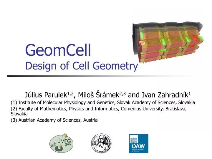
GeomCellDesign of Cell Geometry Július Parulek1,2, Miloš Šrámek2,3 and Ivan Zahradník1 (1) Institute of Molecular Physiology and Genetics, Slovak Academy of Sciences, Slovakia (2) Faculty of Mathematics, Physics and Informatics, Comenius University, Bratislava, Slovakia (3) Austrian Academy of Sciences, Austria
Overview • Introduction to GeomCell • Previous work on geometrical modeling of muscle cells • Representation of mitochondrial shape • GeomCell Implementation • Conclusions and future work
GeomCell - Intro • An environment for virtual cell modeling based on a precise geometric background • possibilities of modern computer graphics and computer hardware to represent a virtual micro-world of cells • first building blocks for representation of a static cell geometry • models of skeletal muscle tissues • idealized models rather than exact reconstruction
Muscle Cell Organelles (Courtesy of Dr. Novotová)
Muscle Cell Organelles Laongitudinal axis Transversal axis (Courtesy of Dr. Novotová)
Input Data - EM images Volume and surface density (stereology) Shape (morphology) Sizes (morphometry) (Courtesy of Dr. Novotová)
Cell Model Organelles • Desired subset of cell organelles: • Myofibrils • Sarcolemma • Sarcoplasmic reticulum • Mitochondria • T-tubules 2 3 1 5 4
f(x) = 0 XISL Implicit Objects f(x) > 0 f(x) < 0 • XISL – Implicit modeling environment • XML based modeling language • C++ library, tools (conversion, rendering, ...)
Cross-sectionalGraphs • Produce carrier skeletons for all virtual organelles • Directly used in modeling of myofibrils • Thin and long cylindrical objects • cross-sectional graphs (c-graph) in a system of parallel planes
Cross-sectionalGraphs(cont.) Minimal distance specification c-graph Real EM images 2D implicit shapes Quadratic interpolation of the 2D shapes
Extended Interpolation • Spatial warp metamorphosis utilized in sarcoplasmic reticulum (SR) modeling • two compartments:terminal cisterns of the SR (A) and Longitudinal SR (B) • skeleton:a set of seed (C) points distributed in a system of cross-sectional planes A B C
Mitochondrial Shape • Elliptically shaped and prolonged organelles of irregular smoothforms and variable sizes • implicit sweep objects
Components of Sweep Objects • A 2D sweep primitive (template) and a 3D sweep trajectory Sweep object 3D trajectory 2D template
Sweep Components for Mitchondria • Template defined as 2D implicit ellipsoid withvariable dimensions • Trajectory as quadratic B-spline
Method Overview • Transformation (MS) maps the 2D template along the curve using so-called reference frames (RF) • Rotation of RF around C’(t) fe
Method Overview (cont.) • Estimate all curve points NP(x)(parameters si), for which x lies in the template planes • Resultant function
Problem of Parameter Estimation • Analytical solution (a, param t) of a general curve trajectory is rarely possible • for instance: cubic spline (C(t) is degree 3) - the polynomial (a) is degree 5 • Solution in using quadratic curves • analytical solution (a)
End Caps • Union with two implicit semi-ellipsoids at both ends
Model Generation • Fully automatic, guided by Model Description Language (MDL) • basic cell dimensions • c-graph distribution • organelle’s geometric parameters specified in a probabilistic sense • Quantification, visualization, …
GeomCell Implementation • Computationally intensive tasks • generation of large number of models • evaluation of volume and surface areas of organelles • model visualization • High throughput computing required • Utilization of a grid environment • retrieval of cell models using metadata (morphological and stereological data, images, MDL spec.,…) • eased with a GUI portal
Conclusions and Future Work • System for cell model generation, quantification, visualization and conversion implemented in Grid environment - GeomCell • Organelle’s behavior • add physical layer to all objects • growth, deformations, cell contraction, … • Pathological cells
Thank You for Your Attention Homepage: www.sccg.sk/~parulek Cell modeling project: www.sccg.sk/~parulek/cell Grid implementation: http://cvs.ui.sav.sk/twiki/bin/view/EGEE/ /GeomCellInEGEE-MuscleCellModelingOnTheGrid Visualization of a volumetric format of a cell model