Download
1 / 127
1.3k likes | 1.48k Views
Immune System. Innate – (Nonspecific) always ready Barriers of skin and mucosae Antimicrobial proteins, phagocytes and other cells Adaptive – (Specific) Attack specific microbes, but slower response. Innate Defenses. I. Surface barriers: skin & mucosae

E N D
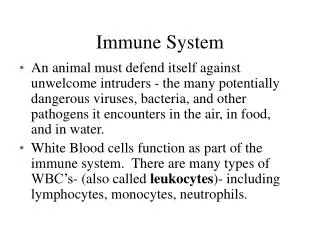
Immune System • Innate – (Nonspecific) always ready • Barriers of skin and mucosae • Antimicrobial proteins, phagocytes and other cells • Adaptive – (Specific) • Attack specific microbes, but slower response
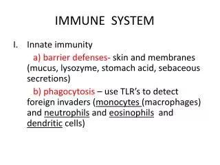
Innate Defenses I. Surface barriers: skin & mucosae • Acidic skin secretions and sebum, vaginal secretions • Stomach acid secretions and enzymes • Saliva and lacrimal secretions w/ lysozyme • Sticky mucus in digestive and respiratory secretions
Innate Defenses cont. II. Internal Defenses: Cells and Chemicals • Phagocytes • NK Cells • Inflammation • Antimicrobial Proteins • Fever
Immunity: Two Intrinsic Defense Systems III. Adaptive (specific) defense system • Third line of defense – mounts attack against particular foreign substances • Takes longer to react than the innate system • Works in conjunction with the innate system
Innate and Adaptive Defenses Figure 21.1
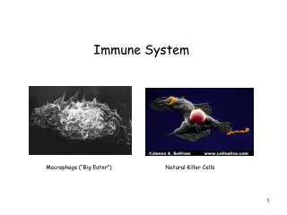
Phagocytes • Macrophages are the chief phagocytic cells • Free macrophages wander throughout a region in search of cellular debris • Kupffer cells (liver) and microglia (brain) are fixed macrophages Figure 21.2a
Phagocytes • Monocytes turn into macrophages • Free – alveolar • Fixed – Kupffer cells • Neutrophils - become phagocytic when encountering infectious material • Eosinophils - are weakly phagocytic against parasitic worms • Mast cells - bind and ingest a wide range of bacteria
Mechanisms of Action • Microbe adheres to phagocyte • Pseudopods engulf microbe • Phagocytic vesicle fuse to lysosome • Killed and digested microbe creates residual body • Waste removed by exocytosis
Mechanism of Phagocytosis • Microbes adhere to the phagocyte • Pseudopods engulf the particle (antigen) into a phagosome • Phagosomes fuse with a lysosome to form a phagolysosome • Invaders in the phagolysosome are digested by proteolytic enzymes • Indigestible and residual material is removed by exocytosis
Microbe adheres to phagocyte. 1 Phagocyte forms pseudopods that eventually engulf the particle. 2 Phagocytic vesicle containing antigen (phagosome). Lysosome Phagocytic vesicle is fused with a lysosome. 3 Phagolysosome Microbe in fused vesicle is killed and digested by lysosomal enzymes within the phagolysosome, leaving a residual body. 4 Acid hydrolase enzymes Residual body Indigestible and residual material is removed by exocytosis. 5 (b) Figure 21.2b
Natural Killer (NK) Cells • Can lyse and kill cancer cells and virus-infected cells • Are a small, distinct group of large granular lymphocytes • React nonspecifically and eliminate cancerous and virus-infected cells • Kill their target cells by releasing perforins and other cytolytic chemicals • Secrete potent chemicals that enhance the inflammatory response
Inflammation • The tissue response to injury • Prevents spread of agents • Disposes of cell debris and pathogens • Sets the stage for repair • Chemical “alarm” – release into extracellular fluid • Vasodilation & increased vascular permeability • Toll-Like Receptors or TLRs - macrophages and boundary cells that are “triggers” in respiratory and GI epithelium • Cytokines released following mast cell histamine (TLR signal) • Kinins • Prostaglandins • Complement • Leukotrienes • All of which cause BV dilation (hyperemia = redness)
Hallmarks of Inflammation • Redness – erythema & hyperemia • Heat – due to dilated BVs • Swelling – exudate/edema • Dilutes • O2 and nutrients for repair • Clotting proteins enter • Pain – pressure on localized nerve endings
Phagocyte Mobilization • Leukocytosis – Leukocyte Inducing Factors • Margination – selectins (BV) & integrins (Neutrophil surface) • Diapedesis – WBCs squeeze through endothelial slits • Chemotaxis – signals to other WBCs from neutrophils
Antimicrobial Proteins 1) Interferons (IFNs) – secreted by viral infected cells – to interfere with viral replication in other cells – nonspecific protection against all viruses • Interferon stimulates the neighboring cells to activate genes for PKR (an antiviral protein) • PKR nonspecifically blocks viral reproduction in the neighboring cell
Antimicrobial Proteins Types of Interferon: (some can be genetically engineered for therapeutic use) • Alpha - from leukocytes except lymphocytes (Tx: use against HPV and possibly hepatitis C) • Beta - helps reduce inflammation from fibroblasts (Tx: MS) • Gamma - from lymphocytes • Also activate macrophages and mobilize NK cells which gives IFNs a mechanism to upset cancer cells as well
Antimicrobial Proteins 2) Complement • 20 different plasma proteins, include: C1 through C9, factors B, D, and P, and regulatory proteins • C-Reactive Protein (CRP) • Producer by liver – marker for inflammation • Binds to pathogens to “mark for destruction” in a process called opsonization • Two pathways • Classical • Alternative
Complement • Amplifies all aspects of the inflammatory response • Kills bacteria and certain other cell types (our cells are immune to complement) • Enhances the effectiveness of both nonspecific and specific defenses
Complement Pathways • Complement can be activated by two pathways: classical and alternative • Classical pathway is linked to the immune system • Depends on the binding of antibodies to invading organisms • Subsequent binding of C1 to the antigen-antibody complexes (complement fixation) • Alternative pathway is triggered by interaction among factors B, D, and P, and polysaccharide molecules present on microorganisms
Complement Pathways • Each pathway involves a cascade in which complement proteins are activated in a sequence where each step catalyzes the next • Both pathways converge on C3, which cleaves into C3a and C3b
Complement Pathways • C3b initiates formation of a membrane attack complex (MAC) • MAC causes cell lysis by interfering with a cell’s ability to eject Ca2+ • C3b also causes opsonization, and C3a causes inflammation
Fever • Natural body response to infection • Some is good – too much is not • Denatures proteins/enzymes • Reset internal thermostat above 36.2o C • Caused by chemical released called pyrogens secreted by macrophages and leukocytes • Results in sequestered Fe and Zn needed by bacteria for multiplication
Adaptive Defenses • Specific defenses to pathogens • Systemic not just a site • Memory after initial exposure • Two parts; • Humoral – antibody-mediated immunity to: 1)bacteria, 2)bacterial toxins, and 3)free viruses • Cellular – cell-mediated immunity to: 1)virus or parasite infected cells, 2)cancer cells, and 3)grafts • Direct by lysing • Indirect by ramping up inflammation or other lymphocytes and macrophages
Antigens • Complete – large proteins, pollens, nucleic acids NAs, lipids • Immunogenicity – able to stimulate proliferation of lymphocytes or Ab • Reactivity – ability to react w/ lymphocytes or Ab • Incomplete (Hapten) – small molecules/peptides, nucleotides, hormones – link up w/ body proteins • Found in poison ivy, dander, detergents, & cosmetics • Allergens thus have reactivity but no immunogenicity, so you don’t “immune” to your allergies
Antigenic Determinants • Only certain parts of an entire antigen are immunogenic • Antibodies and activated lymphocytes bind to these antigenic determinants • Most naturally occurring antigens have numerous antigenic determinants that: • Mobilize several different lymphocyte populations • Form different kinds of antibodies against it • Large, chemically simple molecules (e.g., plastics) have little or no immunogenicity
Self-Antigens: MHC Proteins • Millions of possible combinations so no two people are alike (except identical twins) • Class I MHC on virtually all body cells – displayed peptides that come from: normal protein recycling in the cell or from displayed fragments of infective organisms within the cell • Class II MHC on certain cells that act in immune response – come from outside the cell
Cells of Adaptive Immunity • Lymphocytes: T-cells & B-cells • Positive selection – self recognition • Negative selection – self tolerance • Immunocompetent in Thymus (T) or Bone Marrow (B) • Genetically preprogrammed to respond to antigens (Ag) • Antigen-Presenting Cells: Dendritic cells, Langerhans cells, macrophages, & activated B-cells • Engulf and display Ag fragments
T Cells • T cells mature in the thymus under negative and positive selection pressures • Negative selection – eliminates T cells that are strongly anti-self • Positive selection – selects T cells with a weak response to self-antigens, which thus become both immunocompetent and self-tolerant
B Cells • B cells become immunocompetent and self-tolerant in bone marrow • Some self-reactive B cells are inactivated (anergy) while others are killed • Other B cells undergo receptor editing in which there is a rearrangement of their receptors
Immunocompetent B or T cells • Display a unique type of receptor that responds to a distinct antigen • Become immunocompetent before they encounter antigens they may later attack • Are exported to secondary lymphoid tissue where encounters with antigens occur • Mature into fully functional antigen-activated cells upon binding with their recognized antigen • It is genes, not antigens, that determine which foreign substances our immune system will recognize and resist
Antigen-Presenting Cells (APCs) • Major rolls in immunity are: • To engulf foreign particles • To present fragments of antigens on their own surfaces, to be recognized by T cells • Major APCs are dendritic cells (DCs), macrophages, and activated B cells • The major initiators of adaptive immunity are DCs, which migrate to the lymph nodes and secondary lymphoid organs, and present antigens to T and B cells
Macrophages and Dendritic Cells • Secrete soluble proteins that activate T cells • Activated T cells in turn release chemicals that: • Rev up the maturation and mobilization of DCs • Prod macrophages to become activated macrophages, which are insatiable phagocytes that secrete bactericidal chemicals
Adaptive Immunity: Summary • Two-fisted defensive system that uses lymphocytes, APCs, and specific molecules to identify and destroy nonself particles • Its response depends upon the ability of its cells to: • Recognize foreign substances (antigens) by binding to them • Communicate with one another so that the whole system mounts a response specific to those antigens
Humoral Immune Response • Clonal selection & differentiation of B-cells or the Primary Immune Response: 3-6d lag • Most clones become plasma cells (make 2000 molecules of Ab/sec!) – last 4-5 days • Immunologic memory – long-lived or Secondary Immune Response: only hours lag and peaks in 2-3d
Active & Passive Immunity • Active (acquired or developed/“challenged”) • Natural – get the disease/infection • Artificial – vaccine for the disease • Passive (transferred no “challenge”) • Natural – maternal Ab • Artificial – antisera, antivenom, antitoxin (rabies, botulism, tetanus)
Ab Structure • 4 looping polypeptide chains “T” or “Y” • 2 Heavy Chain - identical 400 AA chains • 2 Light Chain – identical half as long as heavy • Variable (Ag-binding site) and Constant regions • Classes of Ab – determined by Constant region • IgM, IgA, IgG, IgD, IgE • Body can make over a billion different Ab!
IgD – attaches to B-cells and acts as Ag receptor to activate B-cells • IgM – pentamer circulates in plasma & 1st Ig released by plasma cells in 1o response & seen in active infections, while the monomer is a B-cell receptor • IgG – most prevalent circulating Ab against bacteria, viruses & toxins both 1o & 2o, can cross the placenta • IgA – secretory Ab of mucus in digestive, respiratory, sweat, milk • IgE – bound to mast cells & basophils in allergic reactions and only during allergic reactions will it be in plasma
Basic Antibody Structure Figure 21.13a
Antibody Structure • C regions form the stem of the Y-shaped antibody and: • Determine the class of the antibody • Serve common functions in all antibodies • Dictate the cells and chemicals that the antibody can bind to • Determine how the antibody class will function in elimination of antigens