Download

1 / 96
1.22k likes | 2.53k Views
Vital Signs. Module 9. Temperature. Body Temperature. Body temperature is the balance between heat produced in the body and heat loss from the body . Body Temperatures.

E N D
Vital Signs Module 9
Body Temperature • Body temperature is the balance between heat produced in the body and heat loss from the body.
Body Temperatures • Core Temperature – temperature of the deep tissues of the body such as abdominal or pelvic cavities. It is relatively constant • Surface Temperature – temperature of the skin and subcutaneous tissue. It fluctuates depending on the blood supply to the skin and the amount of heat loss to the external environment.
Thermoregulation • Physiologically • Receptors in the skin, abdomen, and spinal cord send messages to the autonomic nervous system that sends the message to the hypothalamus. • The hypothalamus acts as a central thermostat, receiving input from sensors that detect hot or cold temperatures and initiates body responses mainly in the cardiovascular system via vasoconstriction or vasodilation that decrease heat production and increase heat loss.
Thermoregulation • Behavioral • When an individual perceives he is hot or cold, he changes his behavior such as: moves to the shade or sun, regulates the thermostat, removes extra clothes or puts on sweater.
Four Avenues of Heat Loss • Radiation • Conduction • Evaporation • Convection
Radiation • Transfer of heat from the surface of one object to the surface of another without contact • Blood flows from the core internal organs carrying heat to skin and surface blood vessels. • Amount of heat carried to the surface depends on the extent of vasoconstriction and vasodilation regulated by hypothalamus. • 85% of body heat is loss via radiation to the environment • Usually from the body to a cooler surface
Conduction • Transfer of heat from one object to another with direct contact. • When the warm skin touches a cooler object, the heat is loss • Increase amount loss by applying a ice pack, bathing with cool water • Decrease amount of heat loss by applying blankets • Gains heat via conduction when contact is made with a warmer object
Evaporation • Transfer of heat when a liquid is changed to a gas. • When body temperature rises, hypothalamus signals the sweat glands to release sweat. Sweat evaporates from the skin, resulting in heat loss.
Convection • Transfer of heat away by air movements. • Air currents carry away the heat • An electric fan promotes heat loss through convection
Heat Conservation • Vasoconstriction • Decreases the amount of blood that reaches the surface and thus is not lost.
Heat Production • Increase Basal Metabolism Rate • Movement / exercise • Shivering
Factors that Affect Temperature • Age • Activity / Exercise / Sleep • Hormones • Stress • Environment • Medication • Illness
Ranges of Temperature • Children: 98.60 - 99.60 F, oral • Adults: 98.6 + /-1 degree F, oral • Older Adults: 97.6 + / -1 degree F, oral
Variations • Rectal temperature is usually 1 degree higher than oral • Axillary temperature is usually 1 degree lower than oral
Terminology • Pyrexia, hyperthermia, fever – an elevation in body temperature • Hyperpyrexia – a very high fever such as: 1050F • Febrile – having a fever; Afebrile – without a fever • Hypothermia – decreased body temperature below 970 F
Interpretation • The nurse needs to look at the relationship of the vital signs to each other, to previous findings, and to other assessment data. • If the temperature is abnormal, the nurse should notify the physician. • Reassess the temperature more often than ordered – nursing intervention / judgment
Sites / Methods • Oral • Rectal • Axillary • Tympanic Temporal
Oral • Most common and convenient • Sublingual pocket very vascular and responds to changes in core temperature • Disadvantage – varies with what the patient has had in their mouth, mouth breathers • Not safe for infants, children, elderly, disoriented, epileptic, or unconscious
Rectal • Most reliable • Disadvantage– unpleasant for the patient and inconvenient to gain access to the site • Does not respond to body temp changes • Cannot be used in rectal surgery, or stool in bowel
Axillary • Safest, Noninvasive, Easily accessible • Does not always give a true reading of core temperature • Used for infants, children, patients with oral problems or mouth breathers, and irrational patients.
Tympanic • Utilizes the ear which is readily accessible, minimal discomfort, and reflects core temperature • Disadvantage – cost of the equipment and inaccurate reading because of incorrect positioning of the instrument.
Temporal • One of the newer methods for evaluating a person’s temperature is the temporal area of the forehead. • The temporal artery is a branch of the internal carotid artery, but courses within a millimeter or two of the skin's surface over the lateral forehead and is readily accessible.
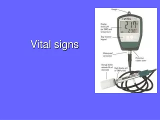
Electronic Probe • Battery operated unit with an attached heat sensitive probe and disposable probe cover. • More rapid reading • Can be used in all methods of assessing the temperature
Tympanic Thermometer • Infrared light reflectance thermometer determines the temperature of the tympanic membrane by measuring heat radiated as infrared energy from that site • Tympanic membrane and hypothalamus share the same vasculature—reflects core temp • Accuracy depends on placement of the device
Temporal Artery Thermometer Temporal Scanner • Designed specifically to be completely non-invasive • Scanner captures naturally emitted infrared heat from the arterial blood supply, locking in the highest temperature it senses
Chemical-in-glass • Measures the temperature by movement of chemical through the calibrated glass. • Must be shaken down to 950before taking a reading. • Disadvantage – must follow hazard precautions if it should break.
Taking a Temperature Oral • Shakes the thermometer to get below 960 or inserts into probe cover • Places in posterior sublingual pocket • Remain in mouth for 3 - 5 minutes • Remove and wipe from stem to bulb (clean to dirty) • Reads at eye level, rotates slowly until mercury visible and reads to nearest tenth of degree • Shakes the thermometer down, cleans with soapy cold water, or pushes ejection button to release probe cover
Taking a Temperature Rectal • Preparation: same • Places the patient in Sim’s position and drape • Apply gloves • Prepares thermometer- lubricates tip
Rectal • Dominant hand hold thermometer, other hand, separate buttocks to expose anus • Instruct patient to take a deep breath. Insert the thermometer gently into the anus: infant- ½ inch, adult 1 ½ inches. • Do not force insertion • Hold in place for 3-5 minutes • Wipe secretions, and discard tissue, read temp
Taking a Temperature Axillary • Gain access to the axillary area • Make sure area is dry • Place thermometer in the center of axilla. Fold patients arm straight down and place arm across chest • Leave in place 6-10 minutes • Remove, read, Write down
Just letting you know • Easy way to remember how long to take a temperature 5 min. = oral +3 min. = rectal 8 min. = axillary
Taking a Temperature Tympanic • Position patient upright or on their side • Attach probe cover to the unit • Turn head to one side and insert into ear canal • Adult – pull pinna upward and back • Child – pull pinna downward and back • Remove after reading displayed • Remove probe cover and place in storage unit
Taking a Temperature Temporal • With just a light stroke across the temporal artery area of the forehead, an accurate reproducible temperature is measured in about 3 seconds - eliminating any discomfort caused by thermometer inserted into the ear, mouth, or rectum.
Definition • A wave of blood created by contraction of the left ventricle of the heart • Heart is a large pump that forces blood to enter the arteries with each heartbeat, causing pressure pulses or pulse waves
CARDIAC OUTPUT • The heart pumps 4-6 liters each minute. • This volume is the cardiac output
Cardiac Output = SV x HR • Stroke volume- amount of blood that enters the arteries with each ventricle contraction • Normally heart empties about 70% of its volume with each contraction. That is about 70 ml. of blood in an adult • Heart rate- beats per minute
Rate of a Pulse • Rate of the ventricular contractions of the heart • Normal rate if 60 – 90 BPM with an extended range of 50- 100 BPM
Terminology - Rates • Tachycardia • Increased heart rate – over 100 BPM • Usually occur when sympathetic nervous system is stimulated • Bradycardia • Decreased heart rate – below 60 BPM • Usually occur with parasympathetic nervous system is stimulated
Factors that Affect the Pulse • Age • Sex • Exercise • Temperature • Medications • Emotions / Stress • Hemorrhage • Illness • Position
Rhythm • Pattern of the beats and the intervals between the beats. • Equal time should lapse between beats of a normal pulse making a normal rhythm • An irregular rhythm is dysrhythmia
Dysrhythmia • May consist of an early beat, late beat, or missed beat • Irregular rhythm may be random, irregular rhythm or a predictable pattern of irregular beats. • Threatens the heart’s ability to provide adequate cardiac output.
Pulse Deficit • Etiology = A contraction of the heart that fails to transmit a pulse wave to the peripheral pulse sites • Assess by apical – radial pulse simultaneously for one minute, compare • Difference between the apical and radial pulse rates is the pulse deficit.
Pulse Deficit • Nurse A takes the apical pulse and gets a rate of 88. • Nurse B takes the radial pulse and gets a rate of 76. • The pulse deficit is 12. 88 – 76 = 12
Strength • Strength of the pulse reflects the volume of blood ejected against the arterial wall with each heartbeat. • Usually the volume / strength is the same with each heartbeat.