Download
1 / 37
410 likes | 511 Views
Blood supply of the Heart & Conduction System. Dr. Nabil Khouri. Arterial supply of Heart. Right coronary artery Left coronary artery. Introduction:. Coronary arteries - VASAVASORUM arising from aortic sinuses of Valsalva of Ascending aorta Rt CA - from Rt aortic sinus (ant)

E N D
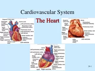
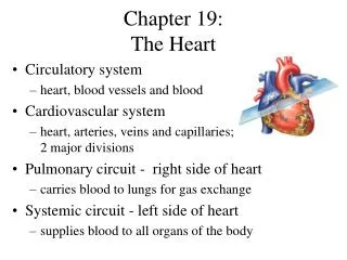
Blood supply of the Heart & Conduction System Dr. Nabil Khouri
Arterial supply of Heart • Right coronary artery • Left coronary artery
Introduction: Coronary arteries - VASAVASORUM arising from aortic sinuses of Valsalva of Ascending aorta Rt CA - from Rt aortic sinus (ant) Lt CA from Lt aortic sinus(left post) Post Aortic sinus - non coronary Max filling of sinuses - in diastole
Right Coronary Artery Conus brs Ventricular brs AV nodal br • Arises from anterior aortic sinus of the Ascending Aorta. • It descends in the right atrioventricular groove. • Near inferior border continuous posteriorly along the atrioventricular groove. • Anastomose with left coronary artery in the posterior interventricular groove.
Branches of Right Coronary Artery • Right conus artery: • Supplies Rt ventricular outflow tract. • Rt marginal branch: • Supply free wall of Rt ventricle.
Branches of Right Coronary Artery • 3. Rt posterolateral branch:Goes back of lt ventricle supply inferior aspect of interventricular septum • Atrial branches:Supply anterior and lateral surface of the right atrium. • One branch supply posterior surface of both right and left atria. • Artery of Sinuatrial Node (60%)
Conus brs Ventricular brs AV nodal br
Crux • Meeting point of • IA groove • Post AV groove • Post IV groove Posteroinferior view Atrioventricular groove (CS) &Post Interventricular groove
Branches of Right Coronary Artery • Posterior interventricular (descending) artery • Runs towards apex in the posterior inter ventricular groove. • Supply right & left ventricles, including its inferior wall. • Supply posterior part of the ventricular septum (Excluding Apex). • Large septal branch Supply Atrioventricular Node.
Branches of Rt coronary Artery Rt conus artery- Annulus of Vieussens SA Nodal br – 60% Ant atrial branches Ant ventr branches Rt Marginal artery: (Largest br) Post ventr branches Post IV br arises near CRUX – 70% br of RCA Post atrial branches AV Nodal artery – 80% Conus brs Ventricular brs AV nodal br
Clinical division of the RCA • Proximal - Ostium to 1st main RV branch • Mid - 1st RV branch to acute marginal branch • Distal - acute margin to the crux
Area of distribution RCA • Rt atrium • Greater part of rt ventricle except area adjoining ant interventricular groove • Small part of lt - ventricle adjoining post interventricular groove • Whole of conducting system of heart except part of lts branch of AV bundle • SA Node –supplied by LCA (40%)
Left Coronary Artery • Larger then Right coronary artery. • Arises from posterior aortic sinus of the Ascending Aorta. • Passes between pulmonary trunk and left auricle. • It enters in the atrioventricular groove and divides into an anterior interventricular branch and a circumflex branch. • Supply greater part of the left Atrium, left ventricle and ventricular septum.
Left Anterior Descending Artery • Coursedown the anterior interventricular groove-usually reaches apex. In 22% of cases does not reach apex. • Branchesseptals and diagonals-supply lateral wall of LV, anterolateral papillary muscle; 37% have median ramus (courses like 1st diagonal). • LADSupplies anterolateral, apex and septum; ~45%-55% of left ventricle.
Left Circumflex Artery • Originfrom distal LMCA. • Course : down distal left AV groove. • Passes around the Apex to enter the posterior interventricular groove & anastomoses with the terminal branches of Right coronary artery. • Branchesobtuse marginal, posterolaterals-supply posterolateral LV, anterolateral papillary muscle. SA node artery-38%. • Supplies15%-25% of LV, unless dominant (supplies 40-50% of LV).
Conus brs Ventricular brs AV nodal br
ClinicaldivisionoftheLAD Proximal - Ostium to 1st major septal perforator Mid - 1st perforator to D2 (90 degree angle) Distal - D2 to end LAD
ClinicaldivisionoftheLCX • Proximal - Ostium to 1st major obtuse marginal branch • Mid - OM1 to OM2 • Distal - OM2 to end LCX
Area of distribution of LCA • Lt atrium • Greater Prt of lt ventricle except post interventricular groove • Small part of rt ventricle adjoining ant interventricular groove • Ant part of interventricular septum • Lt branch of AV bundle
Conducting system of Heart • S-A Node: Right coronary artery (60%) • Left coronary artery (40%) • A-V Node and A-V Bundle: Right coronary artery • Right Bundle branch: Left coronary artery • Left Bundle branch: Right & Left coronary arteries
Cardiacdominance • 85%-Rt dominant coronary artery • 8%- • lt dominant-post descending,posterolateral lt ventricular and AVnodal artery all supplied by terminal portion of lt circumflex coronary artery.rt coronary artery small and supply only rt atrium and rt ventricle • 7%-codominant • RCA-PDA and terminates, circumflex artery-all post Lt ventricular branches
Collateral Circulation-Development • Ischemia and occlusion are the only factors currently recognized to result in significant collateralization. • Usually need very high grade coronary artery occlusion for collaterals to be angiographically apparent.
Venous Drainage of Heart • Coronary Sinus: • Runs in the coronary sulcus (posterior atrioventricular groove). Largest vein of heart About 3 cm long Ends by opening into post wall of rt atrium • Tributaries: • Great cardiac vein • Middle cardiac vein
Coronary Sinus Heart is drained by CS - empties into Rt Atrium. Two set of veins empty directly into Rt Atrium Venae cordis minimi Ant cardiac vein, s/t Rt marginal vein also CS - dilatation of Great Cardiac Vein located in post part of AV groove Opens into Rt atrium b/w IVC and Tricuspid opening guarded by incomplete semicircular “Thebasian valve” Tributaries- all have valves except oblique V of lt atrium
Tributaries of Coronary sinus: 1. Great Cardiac vein Begins near apex of heart; acc. Ant IV A & more proximally cx artery Terminates at lt end of coronary sinus 2. Middle cardiac vein Accompanies Post IV artery and opens at termination of coronary sinus
Cardiac Veins (Sternocostal Surface) Anterior cardiac veins
3. Small Cardiac vein Accompanies rt marginal artery Runs in AV groove to end into rt end of CS May open directly into rt atrium 4. Oblique Vein of Lt Atrium (of Marshall) Runs in the post surface of Lt Atrium and drains into Lt end of Coronary sinus 5. Post Vein of Lt Ventricle Runs on diaphragmatic surface of Lt ventricle and ends in middle of coronary sinus 6. Rt Marginal vein Accompanies Rt Marginal artery and drains into Small Cardiac vein or directly into the Rt Atrium
Oblique Vein of Lt Atrium (of Marshall)
Veins directly emptying into Rt Atrium 1.Ant Cardiac Veins: 3-4 in no .drains the infundibulum of Rt ventricle opens into Rt Atrium through its Ant wall 2. Venae Cordis Minimi/ Thebasian veins Numerous small veins opening into the Post wall of Rt Atrium 3. Small cardiac vein – may open directly into Rt atrium
Contents of Heart grooves 1. Right atrioventricular groove: Right coronary artery Small cardiac vein 2. Left anterior atrioventricular groove: Left coronary artery 3. Left posterior atrioventricular groove: Coronary sinus 4. Anterior interventricular groove: Anterior interventricular artery Great cardiac vein 5. Posterior interventricular groove: Posterior interventricular artery Middle cardiac vein
Venous drainage • Ant cardiac vein and venae cordis minimi opens directly into rt atrium Ant cardiac vein 3-4 small vein running parrelel to one another on ant wall of rt ventricle venae cordis minimi-Thebesian vein or smallest cardiac vein Small vein present in all chambers of heart