Download
1 / 74
870 likes | 1.56k Views
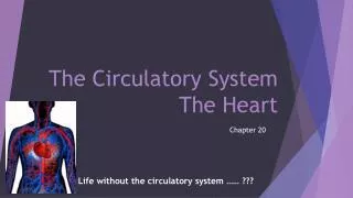
The Circulatory System The Heart. Anatomy & Physiology II Chapter 14. Circulatory System: The Heart. cardiology – the scientific study of the heart and the treatment of its disorders cardiovascular system heart and blood vessels circulatory system heart, blood vessels, and the blood

E N D
The Circulatory SystemThe Heart Anatomy & Physiology II Chapter 14
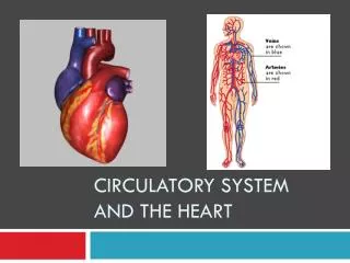
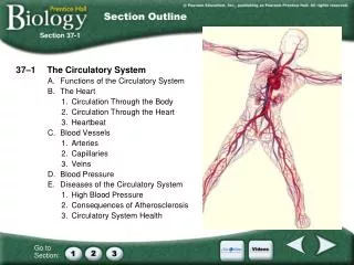
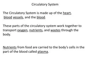
Circulatory System: The Heart • cardiology – the scientific study of the heart and the treatment of its disorders • cardiovascular system • heart and blood vessels • circulatory system • heart, blood vessels, and the blood • major divisions of circulatory system • pulmonary circuit - right side of heart • carries blood to lungs for gas exchange and back to heart • systemic circuit - left side of heart • supplies oxygenated blood to all tissues of the body and returns it to the heart
Circulation and the Heart Circulation • Continuous one-way circuit of the blood vessels • Propelled by heart
Cardiovascular System Circuit CO2 O2 • left side of heart • fully oxygenated blood arrives from lungs via pulmonary veins • blood sent to all organs of the body via aorta • right side of heart • lesser oxygenated blood arrives from inferior and superior vena cava • blood sent to lungs via pulmonary trunk Pulmonary circuit O2-poor, CO2-rich blood O2-rich, CO2-poor blood Systemic circuit CO2 O2
Location of the Heart • Between the lungs • Left of the midline of the body • In mediastinum • Apex pointed toward left
Position, Size, and Shape • heart located in mediastinum, between lungs • base – wide, superior portion of heart, blood vessels attach here • apex - inferior end, tilts to the left, tapers to point • weighs 10 oz. Aorta Pulmonary trunk Superior vena cava Base of heart Right lung Parietal pleura (cut) Pericardial sac (cut) Apex of heart Diaphragm
Heart Wall • epicardium(visceral pericardium) • serous membrane covering heart • coronary blood vessels travel through this layer • endocardium • smooth inner lining of heart and blood vessels • covers the valve surfaces and continuous with endothelium of blood vessels • myocardium • layer of cardiac muscle • muscle spirals around heart which produces wringing motion • fibrous skeleton of the heart - framework of collagenous and elastic fibers • provides structural support and attachment for cardiac muscle and anchor for valve tissue • electrical insulation between atria and ventricles important in timing and coordination of contractile activity
The Heart Wall and Pericardium The serous pericardium covers the heart and lines the fibrous pericardium. ZOOMING IN • Which layer of the heart wall is the thickest?
Pericardium (pericardial sac) • pericardium - double-walled sac that encloses the heart • allows heart to beat without friction, provides room to expand, yet resists excessive expansion • anchored to diaphragm inferiorly and sternum anteriorly • parietal pericardium – outer wall of sac; has two layers • superficial fibrous layer of connective tissue • a deep, thin serous layer • visceral pericardium (epicardium) – heart covering • serous lining of sac turns inward at base of heart to cover the heart surface • pericardial cavity - space inside the pericardial sac (between the visceral and parietal pericardium) filled with pericardial fluid • pericarditis – inflammation of the membranes • painful friction rub with each heartbeat
Pericardium and Heart Wall Pericardial cavityPericardial sac: Fibrous layer Serous layer Epicardium Myocardium Endocardium Epicardium Pericardial sac
Special Features of the Myocardium Cardiac muscles • Are lightly striated (striped) • Have single nucleus cells • Are controlled involuntarily • Have intercalated disks • Have branching muscle fibers
Divisions of the Heart Double pump • Right side pumps O2-poor / CO2-rich blood to the lungs • Pulmonary circuit • Left side pumps oxygenated (CO2-rich) blood to remainder of body • Systemic circuit
Four Chambers • Right atrium • Receives low-oxygen blood returning from body tissue through superior vena cava and inferior vena cava • Left atrium • Receives high-oxygen blood from lungs • Right ventricle • Pumps blood from right atrium to lungs • Left ventricle • Pumps oxygenated blood to body
The Heart as a Double Pump The right side of the heart pumps blood through the pulmonary circuit to the lungs to be oxygenated; the left side of the heart pumps blood through the systemic circuit to all other parts of the body. ZOOMING IN • What vessel carries blood into the systemic circuit?
The Heart and Great Vessels ZOOMING IN • Which heart chamber has the thickest wall?
Blood Flow Through Heart 10 Blood enters right atrium from superior and inferior venae cavae. 1 2 Blood in right atrium flows through right AV valve into right ventricle. Aorta Left pulmonary artery 11 Contraction of right ventricle forces pulmonary valve open. 3 5 5 Blood flows through pulmonary valve into pulmonary trunk. 4 9 Pulmonary trunk Superior vena cava 4 Left pulmonary veins Blood is distributed by right and left pulmonary arteries to the lungs, where it unloads CO2 and loads O2. 5 6 6 Right pulmonary veins Left atrium 1 6 Blood returns from lungs via pulmonary veins to left atrium. Aortic valve 7 3 Left AV (bicuspid) valve 7 Blood in left atrium flows through left AV valve into left ventricle. Right atrium 8 Left ventricle Contraction of left ventricle (simultaneous with step 3 ) forces aortic valve open. 8 2 Right AV (tricuspid) valve 9 Blood flows through aortic valve into ascending aorta. Right ventricle 10 Blood in aorta is distributed to every organ in the body, where it unloads O2 and loads CO2. Inferior vena cava 11 11 Blood returns to heart via venae cavae. blood pathway travels from the right atrium through the body and back to the starting point
Four Valves • Valves ensure a one-way flow of blood through the heart • Atrioventricular valves • Entrance valves • Right AV (tricuspid) and Left AV (bicuspid) • Semilunar valves • Exit valves • Pulmonary and Aortic
Heart Valves • atrioventricular (AV) valves – controls blood flow between atria and ventricles • Triscupid valve has 3 cusps • Bicuspid (miral) valve has 2 cusps • chordae tendineae - cords connect AV valves to papillary muscles on floor of ventricles • prevent AV valves from flipping inside out or bulging into the atria when the ventricles contract • semilunar valves - control flow into great arteries – open and close because of blood flow and pressure • pulmonary semilunar valve - in opening between right ventricle and pulmonary trunk • aortic semilunar valve in opening between left ventricle and aorta
Heart Valves Left AV (bicuspid) valve Right AV (tricuspid) valve Fibrous skeleton Openings to coronary arteries Aortic valve Pulmonary valve
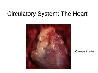
Blood Supply to the Myocardium Coronary circulation • Right coronary artery • Left coronary artery • Coronary sinus
Coronary Circulation • 5% of blood pumped by heart is pumped to the heart itself through the coronary circulation to sustain its strenuous workload • 250 ml of blood per minute • needs abundant O2 and nutrients • left coronary artery (LCA) branch off the ascending aorta • anterior interventricular branch • supplies blood both ventricles and anterior two-thirds of the interventricular septum • circumflex branch • supplies left atrium and posterior wall of left ventricle • right coronary artery (RCA) branch off the ascending aorta • supplies right atrium and sinoatrial node (pacemaker) • right marginal branch • supplies lateral aspect of right atrium and ventricle • posterior interventricular branch • supplies posterior walls of ventricles
Coronary Vessels - Posterior Aorta Left pulmonary artery Superior vena cava Right pulmonary artery Left pulmonary veins Right pulmonary veins Left atrium Coronary sulcus Right atrium Coronary sinus Inferior vena cava Fat Posterior interventricular sulcus Left ventricle Apex of heart Right ventricle Posterior view
Opening of coronary arteries in the aortic valve (anterior view). (A) When the left ventricle contracts, the aortic valve opens. The valve cusps prevent filling of the coronary arteries. (B) When the left ventricle relaxes, backflow of blood closes the aortic valve and the coronary arteries fill.
Coronary Blood Flow • blood flow to the heart muscle during ventricular contraction is slowed, unlike the rest of the body • three reasons: • contraction of the myocardium compresses the coronary arteries and obstructs blood flow • opening of the aortic valve flap during ventricular systole covers the openings to the coronary arteries blocking blood flow into them • during ventricular diastole, blood in the aorta surges back toward the heart and into the openings of the coronary arteries • blood flow to the myocardium increases during ventricular relaxation
Angina • angina pectoris – chest pain from partial obstruction of coronary blood flow • pain caused by ischemia of cardiac muscle • obstruction partially blocks blood flow • myocardium shifts to anaerobic fermentation producing lactic acid stimulating pain
Myocardial Infarction (heart attack) • Interruption of blood supply to the heart from a blood clot or fatty deposit (atheroma) • can cause death of cardiac cells within minutes • Some protection from MI is provided by arterial anastomoses which provides an alternative route of blood flow (collateral circulation) within the myocardium
Myocardial Infarction (heart attack) • myocardial infarction (MI) – sudden death of a patch of myocardium resulting from long-term obstruction of coronary circulation • atheroma (blood clot or fatty deposit) often obstruct coronary arteries • cardiac muscle downstream of the blockage dies • heavy pressure or squeezing pain radiating into the left arm • some painless heart attacks may disrupt electrical conduction pathways, lead to fibrillation and cardiac arrest • silent heart attacks occur in diabetics & elderly • MI responsible for about half of all deaths in the United States
Cardiac Conduction System Electrical energy stimulates heart muscle • Nodes • Sinoatrial (SA) node (pacemaker) • Atrioventricular (AV) node • Specialized fibers • Atrioventricular bundle (bundle of His) • Purkinje fibers (conduction myofibers) • The sinoatrial (SA) node, the atrioventricular (AV) node, and specialized fibers conduct the electrical energy that stimulates the heart muscle to contract.
Cardiac Conduction System 1 SA node fires. 2 Excitation spreads through atrial myocardium. Right atrium 2 1 Sinoatrial node (pacemaker) Left atrium 3 AV node fires. 2 Purkinje fibers Excitation spreads down AV bundle. 3 4 Atrioventricular node Bundle branches Purkinje fibers distribute excitation through ventricular myocardium. Atrioventricular bundle 5 4 5 Purkinje fibers
The Conduction Pathway Sinus rhythm • Sinoatrial (SA) node • Atria • Atrioventricular (AV) node • Internodal pathways • Bundle of His • Bundle branches and Purkinje fibers • Ventricles
Function of the Heart Left and right sides of heart work together in cardiac cycle (heartbeat) • Systole (active phase, contraction) • Diastole (resting phase)
Electrocardiogram (ECG or EKG) • composite of all action potentials of nodal and myocardial cells detected, amplified and recorded by electrodes on arms, legs and chest 0.8 second R R +1 PQ segment ST segment T wave Millivolts P wave 0 PR interval Q S QT interval QRS interval –1 Atria contract Ventricles contract Atria contract Ventricles contract
EKG Deflections • P wave • SA node fires, atria depolarize and contract • atrial systole begins 100 msec after SA signal • QRS complex • ventricular depolarization • complex shape of spike due to different thickness and shape of the two ventricles • ST segment - ventricular systole • plateau in myocardial action potential • T wave • ventricular repolarization and relaxation
Normal Electrocardiogram (ECG) 0.8 second R R +1 PQ segment ST segment T wave P wave Millivolts 0 Q PR interval S QT interval QRS interval –1 Atria contract Ventricles contract Atria contract Ventricles contract
Electrical Activity of Myocardium • atrial depolarization begins • atrial depolarization complete (atria contracted) • ventricles begin to depolarize at apex; atria repolarize (atria relaxed) • ventricular depolarization complete (ventricles contracted) • ventricles begin to repolarize at apex • ventricular repolarization complete (ventricles relaxed) Key Wave of depolarization R Wave of repolarization P P Q S 4 1 Ventricular depolarization complete. Atria begin depolarizing. R T P P Q S Ventricular repolarization begins at apexand progresses superiorly. 2 5 Atrial depolarization complete. R R T P P Q Q S Ventricular depolarization begins at apexand progresses superiorly as atria repolarize. Ventricular repolarization complete; heartis ready for the next cycle. 6 3
Diagnostic Value of ECG • abnormalities in conduction pathways • myocardial infarction • nodal damage • heart enlargement • electrolyte and hormone imbalances
ECGs: Normal and Abnormal • abnormalities in conduction pathways • myocardial infarction • heart enlargement • electrolyte and hormone imbalances (a) Sinus rhythm (normal) (b) Nodal rhythm—no SA node activity
Cardiac Cycle • cardiac cycle - one complete contraction and relaxation of all four chambers of the heart • atrial systole (contraction) occurs while ventricles are in diastole (relaxation) • atrial diastole occurs while ventricles in systole • quiescent period all four chambers relaxed at same time • questions to solve – how does pressure affect blood flow? and how are heart sounds produced?
Timing of Cardiac Cycle • in a resting person • atrial systole last about 0.1 sec • ventricular systole about 0.3 sec • quiescent period, when all four chambers are in diastole, 0.4 sec • total duration of the cardiac cycle is therefore 0.8 sec in a heart beating 75 bpm
The Cardiac Cycle ZOOMING IN • When the ventricles contract, what valves close? What valves open?
Ventricular Filling • during diastole, ventricles expand • their pressure drops below that of the atria • AV valves open and blood flows into the ventricles • end-diastolic volume (EDV) – amount of blood contained in each ventricle at the end of ventricular filling • 130 mL of blood
Ventricular Ejection • ejection of blood begins when the ventricular pressure exceeds arterial pressure and forces semilunar valves open • blood spurts out of each ventricle rapidly at first – rapid ejection • then more slowly under reduced pressure – reduced ejection • stroke volume (SV) of about 70 mL of blood is ejected of the 130 mL in each ventricle • ejection fraction of about 54% • as high as 90% in vigorous exercise • end-systolic volume (ESV) – the 60 mL of blood left behind
Overview of Volume Changes end-systolic volume (ESV) 60 ml -passively added to ventricle during atrial diastole +30 ml -added by atrial systole +40 ml total: end-diastolic volume (EDV) 130 ml stroke volume (SV) ejected by ventricular systole -70 ml leaves: end-systolic volume (ESV) 60 ml both ventricles must eject same amount of blood
Unbalanced Ventricular Output Right ventricular output exceeds left ventricular output. 1 2 Pressure backs up. pulmonary edema Fluid accumulates in pulmonary tissue. 3 1 2 3 (a) Pulmonary edema
Unbalanced Ventricular Output Left ventricular output exceeds right ventricular output. 1 Pressure backs up. 2 peripheral edema Fluid accumulates in systemic tissue. 3 1 2 3 (b) Systemic edema
Congestive Heart Failure • congestive heart failure (CHF) - results from the failure of either ventricle to eject blood effectively • usually due to a heart weakened by myocardial infarction, chronic hypertension, valvular insufficiency, or congenital defects in heart structure. • left ventricular failure – blood backs up into the lungs causing pulmonary edema • shortness of breath or sense of suffocation • right ventricular failure – blood backs up in the vena cava causing systemic or generalized edema • enlargement of the liver, ascites (pooling of fluid in abdominal cavity), distension of jugular veins, swelling of the fingers, ankles, and feet • eventually leads to total heart failure
Cardiac Output (CO) • cardiac output (CO) – the amount ejected by ventricle in 1 minute • cardiac reserve – the difference between a person’s maximum and resting CO • increases with fitness, decreaseswith disease • to keep cardiac output constant as we increase in age, the heart rate increases as the stroke volume decreases
Cardiac Output (CO) Calculating cardiac output • Cardiac output (CO) • Stroke volume (SV) • Heart rate (HR) • cardiac output = heart rate x stroke volume • CO = HR x SV • about 4 to 6 L/min at rest • a RBC leaving the left ventricle will arrive back at the left ventricle in about 1 minute • vigorous exercise increases CO to 21 L/min for fit person and up to 35 L/min for world class athlete
Heart Rate • pulse – surge of pressure produced by each heart beat that can be felt by palpating a superficial artery with the fingertips • infants have HR of 120 bpm or more • young adult females avg. 72 - 80 bpm • young adult males avg. 64 to 72 bpm • heart rate rises again in the elderly • tachycardia - resting adult heart rate above 100 bpm • stress, anxiety, drugs, heart disease, or fever • loss of blood or damage to myocardium • bradycardia - resting adult heart rate of less than 60 bpm • in sleep, low body temperature, and endurance trained athletes
Stroke Volume (SV) • the other factor that in cardiac output, besides heart rate, is stroke volume • three variables govern stroke volume: • preload • contractility • afterload • example • increased preload or contractility causes increases stroke volume • increased afterload causes decrease stroke volume