Download
1 / 22
220 likes | 224 Views
DIGESTIVE SYSTEM. Peritoneum: Membrane of the abdominal cavity. Parietal Peritoneum covers the abdominal wall Visceral Peritoneum covers the inner organs Branching from the peritoneum: Lesser omentum —attaches the liver to the lesser curvature of stomach

E N D
Peritoneum: Membrane of the abdominal cavity • Parietal Peritoneum covers the abdominal wall • Visceral Peritoneum covers the inner organs Branching from the peritoneum: • Lesser omentum—attaches the liver to the lesser curvature of stomach • Greater omentum: (Apron) Contains fat to insulate, cushion, and protect abdominal organs
The Digestive System Functions • Ingestion—taking in food • Digestion—breaking food down both physically and/or chemically • Absorption—movement of nutrients into the bloodstream • Defecation—rids the body of indigestible waste
Organs of the Digestive System • Two main groups of organs • Alimentary canal (gastrointestinal or GI tract) -- continuous coiled hollow tube from mouth to anus • Accessory digestive organs -- aid in digestion but food does not pass through
Layers of Alimentary Canal Organs • Four layers • Mucosa • Submucosa • Muscularis externa • Serosa
Layers of Alimentary Canal Organs • Mucosa • Innermost, moist membrane , smooth muscle layer • Submucosa • Just beneath the mucosa, contains blood vessels and nerve endings • Muscularisexterna—smooth muscle • Inner circular layer and outer longitudinal layer • Serosa—outer membrane
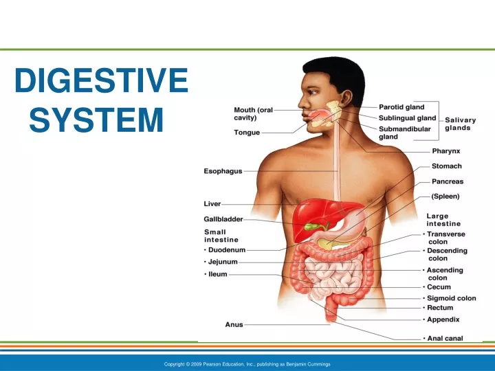
Organs of the Alimentary Canal • Mouth • Pharynx • Esophagus • Stomach • Small intestine • Large intestine • Anus
Mouth (Oral Cavity) Anatomy • Lips (labia)—protect the anterior opening • Cheeks—form the lateral walls • Hard palate—forms the anterior roof • Soft palate—forms the posterior roof • Uvula—fleshy projection of the soft palate
Mouth Physiology • Ingestion • Mastication (chewing) of food • Mixing masticated food with saliva • Initiation of swallowing by the tongue • Allows for the sense of taste • Enzymes from salivary glands are secreted here • Digestion begins here
Pharynx Anatomy (REVIEW) • Nasopharynx—not part of the digestive system • Oropharynx—posterior to oral cavity • Laryngopharynx—below the oropharynx and connected to the esophagus
Pharynx Physiology • Serves as a passageway for air and food • Food is propelled to the esophagus by two muscle layers • Longitudinal inner layer • Circular outer layer • Food movement is by alternating contractions of the muscle layers (peristalsis) http://www.nlm.nih.gov/medlineplus/ency/anatomyvideos/000097.htm
Esophagus Anatomy and Physiology • Anatomy • About 10 inches long • Runs from pharynx to stomach through the diaphragm • Physiology • Conducts food by peristalsis (slow rhythmic squeezing; a wave-like movement of smooth muscles) • Passageway for food only (respiratory system branches off after the pharynx)
Stomach Anatomy • Located on the left side of the abdominal cavity • Food enters at the cardioesophageal sphincter • Contain Rugae—internal folds of the mucosa • Food empties into the small intestine at the pyloric sphincter (valve)
Stomach Physiology • Temporary storage tank for food • Enzymes released here • Digestion occurs here • Hydrochloric Acid produced here • Delivers chyme (processed food) to the small intestine
Subdivisions of the Small Intestine • Duodenum • Attached directly to the stomach • Jejunum • Attaches anteriorly to the duodenum • Ileum • End portion of the small intestine extends from jejunum to large intestine http://nutrition.jbpub.com/resources/animations.cfm?id=1&debug=0
Small Intestine Anatomy • Structural modifications that increase surface area • Microvilli—tiny projections of the plasma membrane (create a brush border appearance) • Villi—fingerlike structures formed by the mucosa • Circular folds (plicae circulares)—deep folds of mucosa and submucosa
Large Intestine • No digestion here • Absorption occurs here • Larger in diameter, but shorter in length, than the small intestine • Frames the internal abdomen
Large Intestine Anatomy • Cecum—saclike first part of the large intestine • Appendix • Accumulation of lymphatic tissue that sometimes becomes inflamed (appendicitis) • Hangs from the cecum • Nursery for important digestive bacteria
Large Intestine Anatomy • No villi present • Goblet cells produce alkaline mucus which lubricates the passage of feces • Muscularis externa layer is reduced to three bands of muscle called teniae coli • These bands cause the wall to pucker into haustra (pocketlike sacs) • Bacteria produce Vitamins
Large Intestine Anatomy • Colon • Ascending—travels up right side of abdomen • Transverse—travels across the abdominal cavity • Descending—travels down the left side • Sigmoid—enters the pelvis • Rectum and anal canal—also in pelvis
Large Intestine Anatomy • Anus—opening of the large intestine • Double sphincter • These sphincters are normally closed except during defecation