Download
1 / 22
220 likes | 419 Views
Perfusion Chamber with Porous Membrane for Cellular-Level Glaucoma Research. Joey Labuz Holly Liske Laura Piechura Kellen Sheedy. Donna Peters, PhD Department of Pathology and Laboratory Medicine William Murphy, PhD Department of Biomedical Engineering. Overview. Project Motivation

E N D
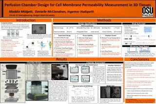
Perfusion Chamber with Porous Membrane for Cellular-Level Glaucoma Research Joey Labuz Holly Liske Laura Piechura Kellen Sheedy Donna Peters, PhD Department of Pathology and Laboratory Medicine William Murphy, PhD Department of Biomedical Engineering
Overview • Project Motivation • Design Specifications • Design Alternatives and Final Design • Design Matrix • Expected Budget • Future Work • Questions
Glaucoma • 2nd leading cause of blindness • 3 million suspected cases in America • Progression: • Elevated intraocular pressure • Optic nerve damage • Loss of vision (National Eye Institute)
Fluid Outflow Pathways normal glaucoma (Modified from Lewis et al., 1999)
Glaucoma Treatment • Current • Prescription eye drops • To decrease production of ocular fluid • Dr. Peters’s research • Injection of ECM peptides • To increase fluid release through the trabecular meshwork
Approach to Research • Isolate cells of the trabecular meshwork • Treat cells with the ECM peptide β-catenin • Induce disassembly of the actin cytoskeleton • Measure fluid flow across the cell layer
Client Motivation • Identify peptides as potential glaucoma therapies • Cellular-level experimentation • Replicates • Cost (Johnson and Tschumper, 1987)
Problem Statement • Cells are more readily available than whole eyes • Device to control fluid flow across cells adhered to membrane • Variable pressure from above and below cells • Secure membrane to membrane holder and pressure device
Design Criteria • Ability to withstand 30 mmHg above and below • Compatibility with various membranes • Ability to perform simultaneous replicates • Integration with existing equipment • Sterile system
Alternative Design One Top Chamber FromSyringe To Transducer = MembraneHolder BottomChamber FromSyringe ToTransducer Stand
Design One Evaluation • Simple design • Interchangeable membrane • No fluid leakage • Difficult to assemble in a sterile setting • Unstable on a laboratory bench
Alternative Design Two Upper Pressure Chamber Membrane Holder = Lower Pressure Chamber
Alternative Design Two From Syringe To Transducer Plan View of Lower Chamber
Design Two Evaluation • Interchangeable membrane • Stable on a laboratory bench • Simple assembly for user • Potential for leakage • Wells cannot be disassembled • Unequal pressure application
Final Design Upper Pressure Chamber = Membrane Holder Lower Pressure Chamber
Final Design Side View To Transducer FromSyringe Front View of Assembled Device
Final Design Evaluation • Magnets provide tight membrane seal • Independent pressure chambers • Minimal user interaction • Upper chamber pressure regulation • Coating of membrane holder
Estimated Budget • Plexiglas (www.professionalplastics.com) • $20 upper chamber • $25 lower chamber • Neodymium magnets (www.amazingmagnets.com) • $20 set of 25 • Small hardware • $30 • Total = $95
Future Work • Coating and sterilizing magnets • Maintaining a constant back pressure • Ordering of materials • Construction • Testing • Fluid flow and measurement • Sealing of membrane http://www.omega.com
References • “Acrylic Sheets: Plexiglas.” Professional Plastics. <http://www.professionalplastics.com> 17 October 2007. • “Back Pressure Regulator.” Plastomatic Valves, Inc. <http://www.plastomatic.com> • “Glaucoma Resource Guide.” National Eye Institute. <http://www.nei.nih.gov/health> 17 October 2007. • Johnson, DH and Tschumper RC. 1987. “Human trabecular meshwork organ culture. A new method.” Investigative Ophthalmology & Visual Science 28: 945-953. • Lewis, Peter R., Phillips, Grant T., and Sassani, Joseph W. April 1, 1999. “Topical therapies for glaucoma: What family physicians need to know.” American Family Physician 59. • “Miniature Back Pressure Regulators.” Omega Process Measurement and Control. <http://www.omega.com> 17 October 2007. • Peters, Donna M. “Use of cell-matrix interactions to treat glaucoma.” PowerPoint presentation. University of Wisconsin-Madison Departments of Pathology and Laboratory Medicine and Opthamology and Visual Sciences. • “Rare Earth Neodymium Magnets.” Amazing Magnets. <http://www.amazingmagnets.com> 17 October 2007. • The Eye Digest. “Glaucoma Treatment.” University of Illinois Eye & Ear Infirmary. 17 June 2007. <http://www.agingeye net/glaucoma/glaucomadrugtreatment.php> 15 October 2007.