Download
1 / 38
420 likes | 605 Views
The Endocrine Pancreas :. Introduction.

E N D
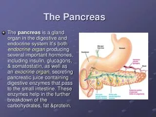
Introduction • The pancreas houses two distinctly different tissues. The bulk of its mass is exocrine tissue and associated ducts, which produce an alkaline fluid loaded with digestive enzymes which is delivered to the small intestine to facilitate digestion of foodstuffs • Scattered throughout the exocrine tissue are several hundred thousand clusters of endocrine cells which produce the hormones Insulin and Glucagon, plus a few other hormones.
Insulin and Glucagon are critical participants in glucose homeostasis and serve as acute regulators of blood glucose concentration. • From a medical perspective, insulin in particular is enormously important - a deficiency in insulin or deficits in insulin responsiveness lead to the disease diabetes mellitus.
The endocrine portion of the pancreas takes the form of many small clusters of cells called islets of Langerhans. • Pancreatic islets house three major cell types, each of which produces a different endocrine product: • Alpha cells (A cells) secrete the hormone glucagon. • Beta cells (B cells) produce insulin and are the most abundant of the islet cells. • Delta cells (D cells) secrete the hormone somatostatin which is also produced by a number of other endocrine cells in the body.
Structure of Insulin • Insulin is protein, composed of two chains held together by disulfide bonds • Proinsulin consists of three domains: an amino-terminal B chain, a carboxy-terminal A chain and a connecting peptide in the middle known as the C peptide
Within the endoplasmic reticulum, proinsulin is exposed to several specific endopeptidases which excise the C peptide, thereby generating the mature form of insulin. • Insulin and free C peptide are packaged in the Golgi into secretory granules which accumulate in the cytoplasm. • When the B cell is appropriately stimulated, insulin is secreted from the cell by exocytosis and diffuses into islet capillary blood. • C peptide is also secreted into blood, but has no known biological activity
Biochemistry of insulin 51 amino acids after cleavage, (86 before) b S S S S Chains S S a C-peptide C-peptide = Connector Peptide LKS
Control of Insulin Secretion • Insulin is secreted in primarily in response to elevated blood concentrations of glucose. • Elevated concentrations of glucose within the B cell ultimately leads to membrane depolarization and an influx of extracellular calcium. • The resulting increase in intracellular calcium is thought to be one of the primary triggers for exocytosis of insulin-containing secretory granules
The normal fasting blood glucose concentration in humans is 80 to 90 mg per 100 ml, associated with very low levels of insulin secretion. • Almost immediately after meals, plasma insulin levels increase dramatically. • Elevated glucose not only simulates insulin secretion, but also synthesis i.e. transcription of the insulin gene and translation of its mRNA
Insulin Release Over Time Insulin Conc. Time 6 min. 90 min. LKS
The Insulin Receptor and Mechanism of Action • The insulin receptor is a tyrosine kinase. • Binding of insulin to the receptor phosphorylate themselves (autophosphorylation), thus activating the catalytic activity of the receptor. • The activated receptor then phosphorylates a number of intracellular proteins, which is insulin receptor substrate 1 or IRS-1, which in turn generate a biological responsethat ultimately mediate insulin's effects.
Peripheral Uptake of Glucose • Glucose cannot diffuse across cell membranes • Tissue uptake is by facilitated transport • *In some tissues insulin regulates the number of membrane transporters
Glucose Transport • GLUT 1 and GLUT 3 mediate basal glucose uptake in most tissues including brain, nerves, and red blood cells. Their high affinities for glucoseensure glucose entry even during periods of relative hypoglycemia. • GLUT 2, a low-affinity transporter, is in hepatocytesand beta-islet cells. After a meal, portal blood from the intestine is rich in glucose. GLUT 2 captures the excess glucose primarily for storage. When the glucose concentration drops below the Km for the transporter, much of the remainder leaves the liver and enters the peripheral circulation. • GLUT 4 is in adipose tissue and muscle and responds to the glucose concentration in peripheral blood. The rate of glucose transport in these two tissues is increased by insulin.
Insulin Actions 1 • Liver Cells • Store Glucose as Glycogen, reduce blood Glucose concentrations • Glycogen synthesis from glucose • Increase glucose transport into cells • Increase Protein Synthesis • Increase amino acid transport into cells • Positive Nitrogen Balance LKS
Insulin Actions 2 • Muscle Cell Actions • Increase glucose uptake • Increase amino acid uptake • Increase protein synthesis (enzymes & structural) • Decrease protein degradation • Increase fatty acid uptake as needed • Increase muscle glycogen synthesis LKS
Insulin Actions 3 • Adipose Cell Actions • Increase protein synthesis ( for lipogenesis) • Increase glucose uptake into cells • Increase lipid formation (lipogenesis) • Decrease lipid degradation (lipolysis) LKS
Effects of insulin on carbohydrate metabolism • Insulin increases the uptake of glucose and its metabolism in muscle and fat. • Insulin increases glycogen synthesis in liver and muscle • Effects of insulin on protein metabolism • Insulin increases amino acid uptake by muscle cells • Insulin increases protein synthesis, and decreases protein breakdown
METABOLIC ACTIONS OF INSULIN • Effects of Insulin on Fat Metabolism • Increase lipid formation (lipogenesis) • Decrease lipid degradation (lipolysis) • Increase lipoprotein lipase activity • Decrease hormone sensitive lipase activity • Insulin Effects on Potassium • Insulin pumps K+ into cells • This K+-lowering action of insulin is used to treat acute, life-threatening hyperkalemia
Tissues that require insulin for glucose uptake are: • Adipose tissue • Resting skeletal muscle • In exercise: glucose can enter exercising muscle without the aid of insulin
Tissues in which glucose uptake is not affected by insulin are: • Nervous tissue • Kidney tubules • Intestinal mucosa • Red blood cells and b-cells of pancreas • Insulin accelerates, but is not required, for glucose uptake by the liver
Diabetes mellitus • Insufficient production of insulin (either absolutely or relative to the body's needs), or the inability of cells to use insulin leads to hyperglycemia and diabetes mellitus. • This latter condition affects mostly the cells of muscle and fat tissues, and results in a condition known as "insulin resistance.“ • This is the primary problem in type 2 diabetes. • The absolute lack of insulin, usually secondary to a destructive process in the pancreas, is the particular disorder in type 1 diabetes. • Only approximately 10% of the patients with diabetes mellitus have type 1 diabetes and the remaining 90% have type 2 diabetes mellitus
Normal fasting plasma glucose levels are less than 110 milligrams per deciliter (mg/dl). • Fasting plasma glucose levels of more than 126 mg/dl on two or more tests on different days indicate diabetes. • A random blood glucose test can also be used to diagnose diabetes. • A random blood glucose level of 200 mg/dl or higher indicates diabetes, but it must be reconfirmed on another day with a fasting plasma glucose or an oral glucose tolerance test.
Glucagon Physiology 1 • Produced in a cells of pancreas • 29 amino acid linear molecule • Stimuli • 1 stimulus is a fall ( ) in plasma glucose • 2 stimulus is rise in gut glucose • 3 stimulus is rise ( ) in plasma amino acids • Circulation via portal blood to Liver LKS
ACTIONS OF GLUCAGON • The primary target for glucagon action is the liver • Note: Skeletal muscle is not a target tissue for glucagon Specific Actions of Glucagon on the Liver • Increases liver glycogenolysis • Increase liver gluconeogenesis • Increases lipolysis in the liver
Effects of Glucagon on Intermediary Metabolism • Carbohydrate Metabolism • Stimulation of glycogenolysis • Inhibition of glycogen synthesis • Stimulation of gluconeogenesis • Inhibition of glycolysis
Lipid Metabolism • Stimulation of lipolysis - Adipose • Stimulation of ketogenesis - Liver • Inhibition of triglyceride synthesis • Protein metabolism • Stimulation of proteolysis