Download
1 / 45
520 likes | 619 Views
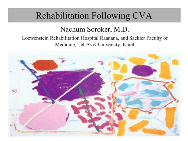
Stroke: An Overview . 台北榮民總醫院 神經醫學中心 神經血管科 許立奇 醫師. What Is Stroke ?. A stroke occurs when blood flow to the brain is interrupted by a blocked or burst blood vessel. Definition of Stroke.

E N D
Stroke: An Overview 台北榮民總醫院 神經醫學中心 神經血管科 許立奇 醫師
What Is Stroke ? A stroke occurs when blood flow to the brain is interrupted by a blocked or burst blood vessel.
Definition of Stroke • Stroke (Cerebrovascular accident, CVA): rapidly developing clinical signs of focal or global disturbance of cerebral function, with symptoms lasting 24 hours or longer, or leading to death, with no apparent cause other than a vascular origin WHO, 1976 • Stroke definition by time course: • Transient ischemia attack (TIA): ischemic events < 24 hours without apparent permanent neurological deficits • Stoke in evolution: progressive neurological deficits over time suggesting a widening of the area of ischemia • Completed stroke: ischemic event with persisted deficit
Stroke Subtypes Ischemic Stroke (83%) Hemorrhagic Stroke (17%) Atherothrombotic Cerebrovascular Disease (20%) Intracerebral Hemorrhage(59%) Cryptogenic and Other KnownCause (30%) Subarachnoid Hemorrhage (41%) Embolism (20%) Lacunar (25%) Small vessel disease Albers GW, et al. Chest. 1998;114:683S-698S. Rosamond WD, et al. Stroke. 1999;30:736-743.
Epidemiology ( I ): Global Burden • 15 million nonfatal stroke each year in the world • Second leading cause of death: 5 million each year • Major cause of permanent disability: another 5 million each year • Risk of stroke: age- and sex-dependent • Incidence: varies with geography • 388/100,000 in Russia, 247/100,000 in China to 61/100,000 in Fruili, Italy
Epidemiology ( II ): Taiwan • Thesecondleading cause of death • Incidence: average annual incidence of first-ever stroke in Taiwan aged 36 years old or over is 300/100,000 (CI: 71%, ICH: 22%, SAH: 1%,others: 6%) • Prevalence: 1,642/100,000 (>36 years old)
Pathophysiology of Ischemic Brain Injury • Brain: • 2% of human body’s mass • 20% of cardiac output • Inadequate perfusion: tissue death and functional deficit • Ischemic brain injury: • A series of interlocking thresholds – the “ ischemic thresholds ” • Decrement in regional CBF key pathologic events
Effects of Reduced CBF Infarction Penumbra Ischemia 50 – 55 25 20 15 8 Edema Loss of Na/K+ electrical pump ↑lactate activity failure; ↓ ATP Normal ml/100g/min Cell Death
Pathophysiology of Ischemic Brain Injury Topography of focal ischemia • Flow gradient: heterogeneous regional CBF reduction after focal ischemia • Densely ischemia region surrounded by areas of less severe CBF reduction • Ischemic penumbra: an area of reduced perfusion sufficient to cause potentially reversible clinical deficits but insufficient to cause disrupted ionic homeostasis
Pathogenesis of Ischaemic Stroke Penumbra Infarction
Risk Factors • Importance: • Identifying those at greatest risk for stroke • Providing targets for preventative therapies • Types: • Modifiable • Non-modifiable
Stroke: Non-modifiable Risk factors • Age • Sex • Ethnicity • Prior stroke • Heredity
Hypertension Diabetes Dyslipidemia Atrial fibrillation Other cardiac conditions Cigarette smoke Asymptomatic carotid stenosis Sickle cell disease Postmenopausal hormone therapy Diet and nutrition Physical Inactivity Obesity and body fat distribution Stroke: Well-Documented and Modifiable Risk Factors
Classification of Ischemic Stroke • By vascular territory • Ant. Circulation: carotid arteries • Post. Circulation: VB system • By stroke etiology
Blood Supply to the Brain:Anterior Circulation • Int. Carotid A. • arises from common carotid a. • Branches: anterior cerebral, anterior communicating, middle cerebral, posterior communicating
What Is the Cause of Ischemic Stroke? • Atherothrombosis • Embolus: • Material: Red (fibrin rich) or White (platelet rich) • Source: Cardiac? Aortic? Carotid Artery? • Small artery disease • Hypoperfusion:Hemodynamic • Others:arterial dissection, arteritis, etc.
Ischemic Stroke: Atherothrombosis • Thrombotic • Acute occluding clot • Superimposed on chronic narrowing
Ischemic Stroke: Cerebral Embolism • Embolic • Intravascular material, most often a clot, separates proximally • Flows through arterial system until it occludes distally • Atrial fibrillation
Ischemic Stroke Subtypes: Data from Taiwan Stroke Registry (2010)
Stroke Warning Signs • Sudden weakness or numbness of the face, arm or leg, especially on one side of the body • Sudden confusion, trouble speaking or understanding • Sudden trouble seeing in one or both eyes • Sudden trouble walking, dizziness/vertigo, loss of balance or coordination • Sudden, severe headaches with no known cause (for hemorrhagic stroke)
Carotid territory Amaurosis fugax Dysphasia Hemiparesis Hemi-sensory loss Vertebrobasilar Hemianopia Quadraparesis Cranial N dysfunction Cerebellar syndrome Crossed deficit Loss of consciousness Localization
Laboratory Examinations • Hb, Hcr, thromb, leuc • glu, CRP, SR, CK, CK-MB, creat • APTT, TT-SPA/INR • Electrolytes, osmolarity • Urine analysis • CSF (if needed for differential diagnosis and only after CT scan, if available) • Others, e.g., coagulation survey, homocysteine for young stroke, rheumotology/immunology screening • Cardiac evaluation: ECG, echocardiography
Intracranial atherosclerosis Penetrating artery disease Flow-reducing carotid stenosis Carotid plaque with arteriogenic emboli Atrial fibrillation Aortic arch plaque Valve disease Cardiogenic emboli Left ventricular thrombi Evaluation of the Vascular System Reprinted with permission from Albers GW, et al. Chest. 2001;119:300S-320S.
Stroke Diagnostic Tests • Brain imaging: CT, MR • Cardiac Imaging: TTE, TEE, heart monitoring • Lipid, coagulation testing • Vascular Imaging: Noninvasive • MR angiography (MRA) Intracranial, extracranial • CT angiography (CTA) Intracranial, extracranial • Ultrasound: Carotid, TCD Invasive • Conventional cerebral angiography Image courtesy of Regional Neurosciences Unit, Newcastle General Hospital, Newcastle, UK.
Diagnosis: CT Scan • Distinguishes reliably between haemorrhagic and ischemic stroke • Detects signs of ischemia as early as 2 h after stroke onset • Identifies haemorrhage immediately • Detects acute SAH in 95% of cases • Helps to identify other neurological diseases (e.g. neoplasms)
CT: Cerebral infarction Brain swelling Focal cortical effacement Ventricular compression
Multimodal CT Imaging CT PCT CTA Vessel Status Tissue Status Perfusion Status CT, computed tomography; PCT, positron computed tomography; CTA, computed tomography angiography. Images courtesy of UCLA Stroke Center.
Differential Diagnosis of Stroke • Ischemic stroke Hemorrhage stroke • Craniocerebral / cervical trauma • Meningitis/encephalitis • Intracranial mass • Tumor • Subdural hematoma • Seizure with persistent neurological signs • Migraine with persistent neurological signs • Metabolic • Hyperglycemia (nonketotic hyperosmolar coma) • Hypoglycemia • Post-cardiac arrest ischemia • Drug/narcotic overdose
Diagnosis: MRI (DWI and PWI) Acute Ischemic Stroke • Diffusion-weighted imaging (DWI) : • Detects areas of restricted diffusion of water • Bright-up in acute ischemic stroke • Differentiation between new and old lesions • Perfusion-weighted imaging (PWI): • Detects abnormal tissue perfusion • Diffusion-perfusion mismatch: • Area of penumbra? • Target of thrombolysis
Multimodal MRI Imaging DWI PWI MRA Vessel Status Perfusion Status Tissue Status DWI, diffusion-weighted imaging; PWI, perfusion-weighted imaging; MRA, magnetic resonance angiography. Images courtesy of UCLA Stroke Center.
Diagnosis: Vascular Imaging Carotid Ultrasound Cerebral Angiography
Management of Cerebrovascular Disease: Current Strategies • Treatment of risk factors in large populations • Treatment of highest risk persons • Management of acute stroke • Prevention and treatment of medical and neurological complications • Rehabilitation • Prevention of recurrent stroke
Strategies for Preventing Stroke and Reducing Stroke Disability stroke mortality blood pressure glucose smoking lipids mass popl. strategy acute treatment recurrent stroke First stroke Secondary prevention high risk strategy Rehabilitation hypertension TIA Atrial fibrillation other vascular disease Stroke related disability
Stroke Therapy: Overview • Risk Factors: • Lifestyle modification • Risk factor management • Acute stroke therapy • Prevention of stroke: • Primary prevention • Secondary prevention
Management of Risk Factors • Non-pharmacological intervention: • Life style modification: cessation of smoking, drinking • Exercise, weight reduction • Pharmacological intervention: • DM, HTN, hyperlipidemia, cardiac diseases,
Management: Improved CBF • Prevention: endarterectomy, stenting • Acute management: thrombolytics – medical and mechanical • Targeting endothelial cell functions (ACEI, calcium blocker, statins, etc.) Brain tissue ischemia Cerebral arterial stenosis/occlusion LAA/CE/SVD/others Decreased CBF Cerebral autoregulation (endothelial function etc)
Antithrombotic Therapies to Prevent Ischemic Stroke • Oral anticoagulants • Antiplatelet agents • Aspirin 50-325 mg/day • Ticlopidine 250 mg twice daily • Clopidogrel 75 mg/day • Aspirin (25 mg) plus extended-release dipyridamole (200 mg) twice a day