Download
1 / 133
1.34k likes | 1.35k Views
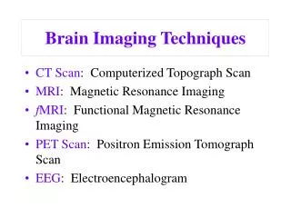
Introduction to Imaging Techniques. Group Work. Answers. Dr Fraser C Millard MA FRCS FRCR. Consultant Radiologist , Warwick Hospital Honorary Associate Clinical Professor, Warwick Medical School. Bowel Obstruction. Question 1. Describe and name the structure shown?. Answer 1.

E N D
Group Work Answers
Dr Fraser C MillardMA FRCS FRCR Consultant Radiologist , Warwick Hospital Honorary Associate Clinical Professor, Warwick Medical School
Question 1 • Describe and name the structure shown?
Answer 1 • Bowel which lies centrally in the abdomen and has thin mucosal folds (valvulae conniventes) which usually form complete lines across the bowel lumen • The small intestine
Question 2 • How has it been demonstrated?
Answer 2 • Small bowel enema • Specially designed tube passed nasally and manoeuvred into the duodeno-jejunal flexure under fluoroscopy • 1 litre of dilute barium infused until a continuous column reaches the terminal ileum
Question 3 • Describe and name the structure shown?
Answer 3 • Bowel which lies peripherally and has thick mucosal folds (haustra) which do not cross the whole bowel lumen • The large intestine
Question 4 • How has it been demonstrated?
Answer 4 • Double contrast barium enema • Full bowel preparation • Barium introduced into the empty colon • Double contrast is achieved by distending the barium coated colon with gas so that the mucosa is seen as a thin white line
Question 5 • What are the symptoms of bowel obstruction?
Answer 5 • Colicky abdominal pain • Small bowel → central (midgut pain) • Large bowel → lower (hindgut pain) • Distension • Nausea & vomiting • Constipation
Question 6 • What is colic?
Answer 6 • Mediated by the autonomic sympathetic nervous system • Associated • Nausea and vomiting • Sweating • Pallor • Severe • Poorly localised and may radiate • Comes in waves • Not relieved by lying still
Question 7 • What are the physical signs in bowel obstruction?
Answer 7 • Scars • Hernias • Abdominal mass • Distension • Visible peristalsis • Increased tinkling bowel sounds
Question 8 • What is the diagnosis and why? • 74 year old gentleman • PC • Several days • Vomiting • Colicky abdominal pain • Bowels not opening & not passing flatus • Abdominal distension
Answer 8 • Small bowel obstruction secondary to a right inguinal hernia
Answer 8 • Obstructed small bowel tends to • Lie centrally in the abdomen • Have thin valvulae conniventes which usually form complete lines across the bowel • Have a diameter of 3-5 cm
Question 9 • What is a hernia?
Answer 9 • A hernia is “the protrusion of an organ or part of an organ through a defect in the wall of the cavity containing it into an abnormal position” • Usually used with reference to the abdomen • Lecture Notes on General Surgery • H Ellis, RY Calne, CJE Watson • Tenth edition 2002 Blackwell Publishing
Question 10 • What is the diagnosis and why? • 69 year old gentleman • PC • 3 day history • Abdominal pain • Distension • Constipation
Answer 10 • Large Bowel Obstruction
Answer 10 • Obstructed large bowel tends to • Lie peripherally • Have thick haustra which do not cross the whole bowel lumen • Have a diameter >5 cm
Question 11 • What examination has been performed to investigate the underlying aetiology and what does it show?
Answer 11 • A contrast enema • An obstructing carcinoma of the sigmoid colon with an apple-core stricture
Question 12 • What examination has been performed in this 81 year old gentleman who has presented with large bowel obstruction and what does it show?
Answer 12 • A CT of the abdomen & pelvis • Large bowel obstruction • Obstructing carcinoma of the descending colon
Question 13 • What advantages does CT have over a contrast enema?
Answer 13 • CT is less invasive and may be better tolerated in an unwell patient • CT has the advantage of providing some staging information • Local spread through the bowel wall • Distant spread • Lymphadenopathy • Liver metastases
Question 14 • What examination has been performed to stage this patient with colonic carcinoma? • What does it show?
Answer 14 • CT thorax abdomen and pelvis • The colonic carcinoma spreads through the bowel wall (T3) • There is spread to the local lymph nodes (N2) • There are metastases to the liver (M1)
Question 15 • How would you approach the question “What are the causes of bloody diarrhoea?”
Answer 15 • VITAMIN • Vascular • Infection • Trauma • Autoimmune • Metabolic • Idiopathic • Neoplasia • Student BMJ “Management Plans” • http://archive.student.bmj.com/issues/08/10/education/364.php
Question 16 • What are the most likely causes in this patient? • 38 year old lady • PC • Acutely ill over 2 weeks • Bloody diarrhoea
Answer 16 • Secretory due to intestinal inflammation • Inflammatory bowel disease • Crohn’s disease • Ulcerative colitis • Infection • Bacterial • Eg campylobacter, bacterial dysentery (Shigella) • Protozoal • Eg amoebic dysentery (Entamoeba histolytica)