Download
1 / 37
420 likes | 506 Views
Adhesion molecules, intercellular connections, epithelial cells. Dr. Zita Puskár EM1.17/09/2018. Levels of body organization. The cell. inside - intracellular, outside - extracellular. What is in the extracellular space?. Extracellular matrix: Fibers Ground substances Water.

E N D
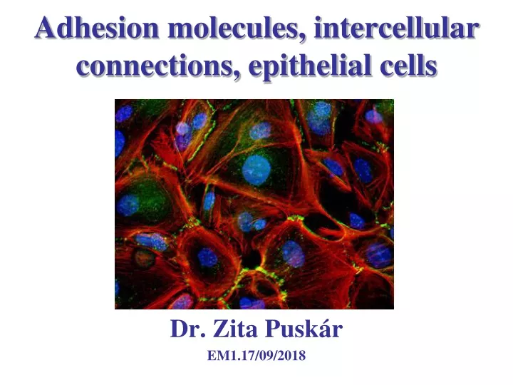
Adhesion molecules, intercellular connections, epithelial cells Dr. Zita Puskár EM1.17/09/2018
The cell inside - intracellular, outside - extracellular
What is in the extracellular space? Extracellular matrix: • Fibers • Ground substances • Water
What is tissue? cell http://www2.optics.rochester.edu/workgroups/cml/opt307/spr06/joe/ Definition: Tissue is a group of cells together with any extracellular secretion that is specialized to perform a specific function. It may contain one type or several types of cells.
The basic tissue types 1. 3. 4. Muscle tissue 2.
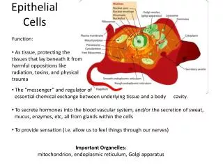
Cells of a tissue Cells (more than one) with common origin and similarity in morphology/shape, composition operation and function Cell FIGURE 9-3 Scanning electron micrograph of the airway surface shows epithelial cells with cilia, possible surface goblet cells dehiscing, and some microvilli. (From Nowell JA, Tyler WS: Scanning electron microscopy of the surface morphology of mammalian lungs, Am Rev Respir Dis 103:313, 1971.)
How is the tissue kept together? by cell adhesion Specific binding of a cell to other cells or to the extracellular matrix (ECM) is called cell adhesion (CA), mediated by interactions between transmembrane glycoproteins called cell adhesion molecules (CAM) Structural elements of CA: (1) CAM, (2) intracellular attachment protein(s)/link protein(s), (3) cytoskeleton Function: development of tissues, cell migration, signal transduction, regulation of gene expression, cell proliferation and cell death, pathological processes (e.g. cancer metastasis)
Adhesion proteins • Common features: • Extracellular domain (interaction with other CAMs (homophilic binding) or ECM (heterophilic binding) • Transmembrane domain • Intracellular domain (interaction with the cytoskeleton)
CAM families Calcium independent Immunoglobulin (Ig) superfamily • weak adhesion • either homophilic or heterophilic e.g. N-CAM (neuronal), V-CAM (vascular) Calcium dependent Cadherins • strong adhesion • homophilic • junctional complexes (adherent junction, link protein: catenin, cytoskeleton: actin filaments) e.g. N-Cadherin (neuronal), E-Cadherin (epithelial), P-Cadherin (placental), desmogleins, and desmocollins, Selectins • weak adhesion • heterophilic e.g. E-selectin (endothelial), L-selectin (leukocyte), P-selectin (platelet) Integrins • cell and ECM • heterophilic • signal transduction e.g. LFA-1, MAC-1
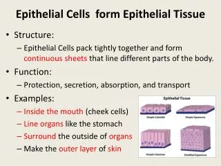
Epithelial Tissue • Features of the epithelial cells: • Closely aggregated polygonal cells • Very little extracellular space • Strong adhesion among the cells • Epithelial tissue is non-vascularized (no bloodvessels) !!! • Functions: • Covering, lining and protecting the surfaces • Absorption (epithels in the intestines) • Secretion (glands) • Contractility (myoepithelial cells) • Specialized sensory function (taste bud, olfactory epithelium)
Simlpe squamous cuboidal columnar Pseudostratified columnar Stratified (Classification is based on the cell shape of the superficial layer) squamous cuboidal columnar Transitional epithelium (urothelium) keratinized non-keratinized
Cell renewal https://www.youtube.com/watch?v=OKosGSm7Ps4 by Walter Jahn
Types of epithelia Surface (Covering) Surface (Lining) Glandular Modified epithelial cells Sensory cells (taste bud, olfactory epithelium)-Neuroepithelium Myoepithel cells Pigment epithelia
Composition of the cytoskeleton Microtubules Microfilamens - actin filaments Filaments Cytoskeleton Intermediate filaments Accessory proteins Motor proteins
Specializations of the apical surface Microvilli Stereocilia Cilia, kinocilia
Junctional complexes gj (a) Occluding junction, zonula occludens (tight junction) (b) adhesive junction, zonula adherens (c) macula adherens (desmosome) (d) hemidesmosome (e) gap junction
General features of junctional complexes Transmembrane adhesion proteins: Connecting cell to cell → homophil binding (occludin, cadherin: Ca2+ dependent adhering protein), Connecting cell to matrix → heterophil binding (integrins) Link proteins– connecting transmembrane adhesion proteins to cytoskeleton (spectrin) Components of the cytoskeleton (actin filaments, intermedier filaments) Types: Homophil-heterophil Ca2+ dependent – Ca2+ independent Strong - weak
Zonula occludens – tight junction Transmembrane proteins: occludin (Ca2+ dependent) claudin, JAM (Junctional Adhesion Protein) Link (anchoring) proteins: Zo1, Zo2, Zo3 Actin filaments Function: separation of the apical and basolateral surfaces, diffusion barrier in the membrane and intercellular space, prevention of the lateral diffusion of membrane components
Zonula adherens Function: • Firm mechanical junction • Encircles the cell • Ca2+ sensitive • Insensitive to osmotic changes • Not a diffusion barrier Actin related (in heart muscle: band like - fascia adherens, in synapse: patch like - punctum adherens)
Macula adherens - desmosome plaque • Related to intermedier filaments (e.g. keratin filaments in epithelia) → form networks • Patch like (patent) • Firm connection (skin, heart muscle) Desmosomal cadherins : desmoglein and desmocloin - together desmoglea Cytoplasmatic plaque: desmoplakin, plaktoglobin
A disease of desmosomes: pemphigus Blistering autoimmune disease in which antibodies form against desmoglein (the transmembrane desmosomal cadherin) and the cells of stratum spinosum are separated from each other (unglued). (Blisters→sores)
Hemidesmosome - Half desmosome This is not a desmosome! Binds the epithelium to the basement membrane Transmembrane proteins – integrins bind to intermedier filaments of the cell and to laminin molecule of basal lamina
Gap junction Channel pore: 1.5 nm • Structural unit: connexon that consists of 6 subunits. The major protein of the subunits: connexin • Small molecules, ions move through the pores • Special signal transduction (cAMP, cGMP) • Opening and closing of the channel is Ca2+ and pH dependent • Appears also in heart muscle, electric synapse gap: 2-4 nm
Basement membrane Function: structural and filtering function, influence cell polarity, regulate cell ploriferation and differentiation, influence cell metabolism and survival , pathway for migration
Transport across epithelia Sodium pumps (Mg2+ activated Na+/K+ ATP-ase) Transcellular transport (fluids and ions) Transcytosis (pinocytotic vesicles)
References: Röhlich Pál: Szövettan, Budapest, 2006 Anthony L. Mescher: Junqueira’s Basic Histology, New York, 2010 Michael Ross and Lynn J. Romrell: Histology, Baltimore, 1989 Geoffrey M. Cooper and Robert E. Hausman: The Cell, A molecular Approach, (ASM, Sinauer), Washington, Sunderland, 2009 Darvas Zsuzsa és László Valéria: Sejtbiológia, Budapest 2005