Download
1 / 20
310 likes | 514 Views
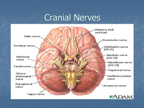
The Vestibulo-cochlear Nerve (Cranial Nerve 8) (Vestibular & Auditory Pathways). OBJECTIVES. At the end of the lecture, the students should be able to: List the nuclei related to vestibular and cochlear nerves in the brain stem. Describe the type and site of each nucleus.

E N D
The Vestibulo-cochlear Nerve (Cranial Nerve 8)(Vestibular & Auditory Pathways)
OBJECTIVES At the end of the lecture, the students should be able to: • List the nuclei related to vestibular and cochlear nerves in the brain stem. • Describe the typeand site of each nucleus. • Describe the vestibular pathways and its main connections. • Describe the auditory pathway and its main connections.
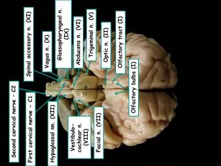
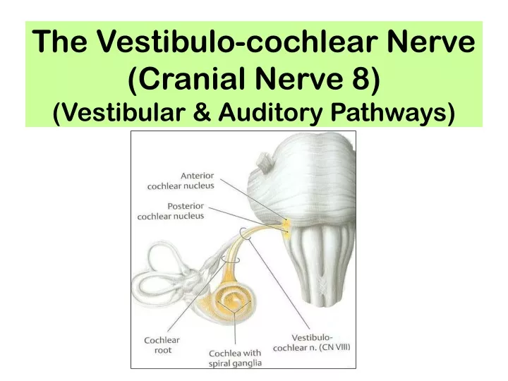
Vestibulo-Cochlear Nerve • Type: Special sensory (SSA) • Conveys impulses from inner ear to nervous system. • Components: • Vestibular part: conveys impulses associated with balance of body (position & movement of the head) • Cochlear part: conveys impulses associated with hearing. • Vestibular & cochlear parts leave the ventral surface of brain stem through the pontomedullarysulcus‘at crebellopontine angle’ (lateral to facial nerve), run laterally in posterior cranial fossaand enter the internal acoustic meatusalong with 7th nerve.
Vestibular Nerve Vestibular nuclei belong to special somatic afferent column in brain stem. • The vestibular nerve fibers make dendritic contact with hair cells of the membranous labyrinth (inner ear). • Their cell bodies (1st order neurons) are located in the vestibular ganglion within the internal auditory meatus. • Their central processes: • Mostly end up in the lateral, medial, inferior and superior vestibular nuclei (2nd order neurons) of the rostral medulla, located beneath the lateral part of the floor of 4th ventricle • Some fibers go to the cerebellum through the inferior cerebellar peduncle 2 1
The efferents from the vestibular nuclei project: To ipsilateralflocculonodular lobe of cerebellum through inferior cerebellar peduncle Bilaterally to ventral posterior nucleus of thalamus, which in turn project to the cerebral cortex. Bilaterally to motor nuclei of cranial nerves through medial longitudinal fasciculus To Motor neurons of the spinal cord as lateral (ipsilateral) & medial vestibulospinal (bilateral) tracts. • Efferentsfrom the vestibular nuclei project to number of other regions for the control of posture, maintenance of equilibrium, co-ordination of head& eye movements and the conscious awareness of vestibular stimulation . 2 1 3 4
Medial Longitudinal Fasciculus • Extends through out the brain stem and formed of both descending & ascending fibers • Projects bilaterally • Has two components: • The ascending component establishes connections with the nuclei of the Occulomotor, Trochlear & Abducent nerves (motor nuclei for extraoccular muscles) for coordination of head & eye movements. • The descending component extends into the spinal cord as the medial vestibulospinal tract.
Vestibulospinal Tracts • Vestibulospinal fibers influence the activity of spinal motor neurons concerned with the control of body posture and balance • Two tracts: lateral & medial • Lateral arises from lateral vestibular (Deiter’s) nucleus, descends ipsilaterally • Medial is the descending part of the medial longitudinal fasciculus, projects bilaterally Lateral
Vestibular Cortex • Located in the lower part of postcentralgyrus (head area). • Responsible for conscious awareness of vestibular sensation.
Auditory Pathway • It is a multisynaptic pathway • There are several locations between medulla and the thalamus where axons may synapse and not all the fibers behave in the same manner. • Representation of cochlea is bilateral at all levels above cochlear nuclei.
Cochlear (Auditory) Nerve Cochlear nuclei belong to special somatic afferent column in brain stem. • The cochlear nerve fibers make dendritic contact with hair cellsof theorgan of Cortiwithin the cochlear duct of the inner ear. • Their cell bodies (1st order neurons)are locatedwithin the cochleain the spiral ganglion. • Their central processes terminate in the dorsal and ventral cochlear nuclei (2nd order neurons), which lie close to the inferior cerebellar peduncle (ICP) Section in Organ of Corti ICP ICP
From the cochlear nuclei, 2nd order neurons, fibres ascend into the pons, where: • Some fibers cross the midline in trapezoid body (1)and terminate in the nucleus of trapezoid bodyor in the contralateralsuperior olivary nucleus (2) • Some fibers run ipsilaterally and terminate in the superior olivary nucleus • From the superior olivary nuclei, ascending fibers comprise the lateral lemniscus (3), which runs through tegmentum of pons and terminate in the inferior colliculus(4) of the mdibrain. 4 4 3 3 5 2 2 1
7 7 Auditory radiation • Some axons within lateral lemniscusterminate in small nucleus of the lateral lemniscus(5) • The inferior colliculi project to medial geniculate nuclei of thalamus(6) • The axons originating in the medial geniculate nucleus (auditory radiation) pass through sublenticular part of the internal capsule to the primary auditory cortex (Brodmann’s areas 41, 42) located in the dorsal surface of the superior temporal gyrus(Heschl’sgyri)(7) 6 6 4 4 5 5 3 3 2 2 1
The region surrounding the primary auditory cortex is known as the auditory association cortex or Wernick’s area (Brodmann’s areas 22) • Wernick’s area is related to recognition and processing of language by the brain
Superior olivary nucleus sends olivocochlear fibers to end in organ of Corti through the vestibulocochlear nerve. These fibers are inhibitory in function and serve to modulate transmission to the cochlear nerve Superior olivary nucleus & the nucleus of the lateral lemniscus establish reflex connections with motor neurons of trigeminal and facial motor nuclei mediating contraction of tensor tympani and stapedius muscles in response to loud noise Inferior colliculi establish reflex connections with motor neurons in the cervical spinal segments (tectospinal tract) for the movement of head and neck in response to auditory stimulation
Clinical Notes • Lesion of vestibulocochlear nerve produces deafness (disturbnce of cochlear nerve functions), tinnitis, vertigo, dizziness, nausea, nystagmus, loss of balance and ataxia (disturbnce of vestibular nerve functions). Acoustic neuroma: a benign tumour of 8th nerve leads to compression of the nerve leading to attacks of dizziness, and profound deafness and ataxia • The representation of cochlea is essentially bilateral at all levels rostral to the cochlear nuclei • Lesions anywhere along the pathway usually have no obvious effect on hearing. • Deafness is essentially only caused by damage to the middle ear, cochlea, or auditory nerve.
SUMMARY • Ganglia related to vestibulocochlear nerve are located in the inner ear. • Vestibular & cochlear nerves pass through internal auditory meatus to cranial cavity, then enter pons at pontocerebellar angle, lateral to facial nerve. • Cochlear & vestibular nuclei are of the special somatic afferent type, and are located in pons & medulla.
SUMMARY • Inferior colliculi,medial geniculatenucleus and finally auditory cortexare stations in cochlear pathway. • Hearing is bilaterally represented. • Vestibular nuclei are connected to: spinal cord (directly or through medial longitudinal fasciculus), to flocculo-nodular lobe of cerebellum and to vestibular area of cerebral cortex.
QUESTION 1 • The third order neurones of auditory pathway are found in: • Mid brain. • Thalamus. • Pons. • Cerebral cortex.
QUESTION 2 • Regarding the vestibular pathway: • The vestibular ganglion is located in the middle ear. • The vestibular nuclei are located in the midbrain. • The vestibular nuclei are connected to the cerebellum. • The vestibulospinal tracts are located in the lateral white column of spinal cord.