Download
1 / 43
430 likes | 435 Views
Learn about the structure and function of the heart, including its chambers, valves, and blood flow. Understand how the heart circulates blood throughout the body and how oxygen and carbon dioxide are exchanged in the lungs.

E N D
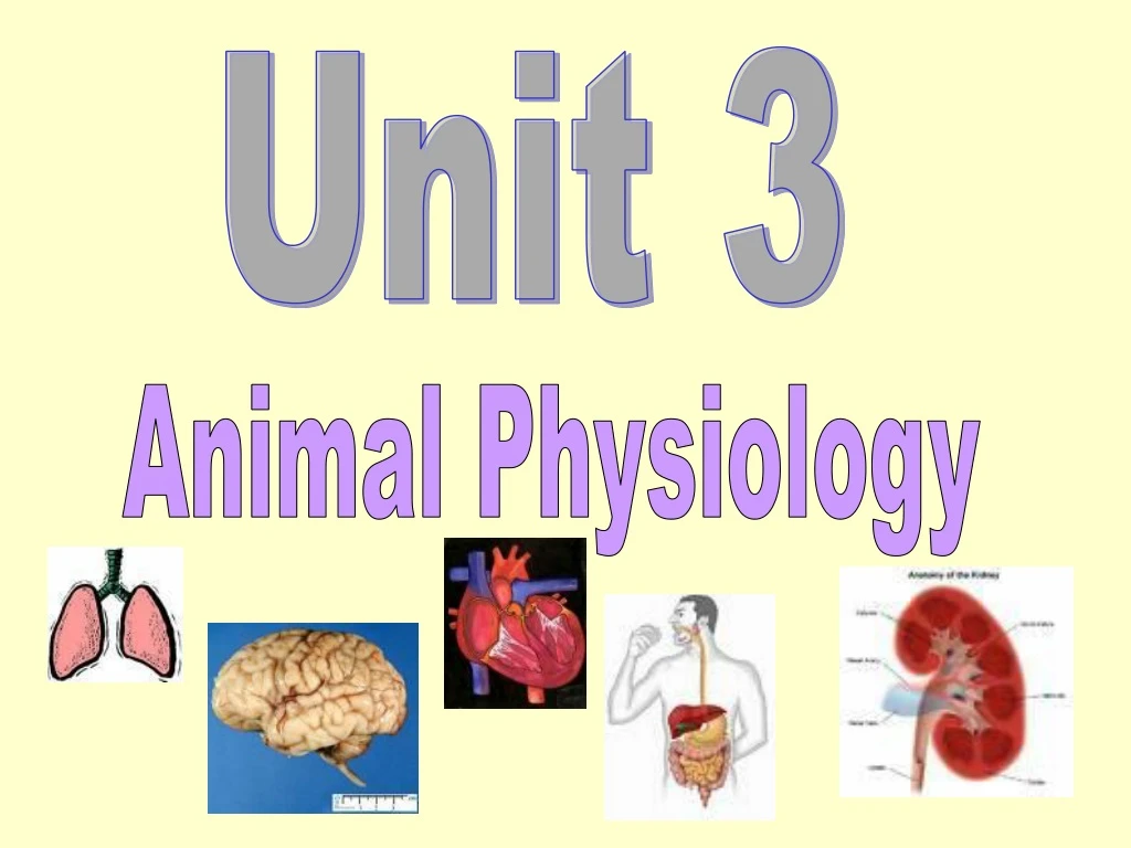
Unit 3 Animal Physiology
Circulation and Gas Exchange The Heart • The heart is a muscular organ that is divided into 4 chambers:- 2 atria and 2 ventricles. (left and right) • The left ventricle wall is very thick and muscular since it has to pump blood all round the body. • The right ventricle wall is less thick since it just pumps blood to the lungs.
The heart has 4 heart valves. • 2 of the valves are between the atria and ventricles. On the left hand side is the bicuspid valve. On the right hand side is thetricuspid valve. • The 2 other heart valves are at the origins of the pulmonary artery and the aorta – these are the semi-lunar valves. • The valves ensure that blood is only able to flow in one direction. They prevent backflow of blood.
Semi lunar valves aorta Pulmonary artery Pulmonary vein Vena cava Left atrium Right atrium Bicuspid valve Tricuspid valve Right ventricle Left ventricle
Since the heart wall is made of muscle it must get its own supply of oxygenated blood. • The heart is supplied by the coronary arteries. (a branch of the aorta). • If a coronary artery becomes blocked, the heart does not get a supply of oxygen and this may result in death of the tissue.
Path of Blood Flow Through the Heart • Blood arrives at the heart via the vena cava. • This blood is low in oxygen (deoxygenated.) • From the vena cava it enters the right atrium then the right ventricle. • It exits the heart by the pulmonary artery where it is carried to the lungs to pick up oxygen. • The blood becomes oxygenated. • From the lungs it goes back to the heart via the pulmonary vein. • It enters the left atrium, then the left ventricle. • The blood leaves the heart in the aorta where it is delivered to the rest of the body.
Head & Body Vena cava Right atrium Pulmonary artery Right ventricle Lungs Pulmonary vein Left atrium Left ventricle Head & Body Aorta
Semi lunar valves aorta Pulmonary artery Pulmonary vein Vena cava Left atrium Right atrium Right ventricle Left ventricle
Circulation & Blood Vessels ARTERIES • Arteries carry blood away from the heart. • Arteries carry oxygenated blood to organs and tissues – (except the pulmonary artery) • Where an artery lies just under the skin the beating of the heart can be felt. Each push of blood is called a pulse. • Artery walls are very thick and muscular since they have to withstand blood travelling at high pressure.
VEINS • Veins carry blood to the heart • Veins carry deoxygenated blood from organs and tissues (except the pulmonary vein) • Veins have valves present to stop the blood flowing backwards.
CAPILLARIES • Arteries split into a network of tiny thin walled vessels called capillaries. • Capillaries are 1 cell thick, they are long, narrow and thin to provide a large surface area. • Capillaries carry food and oxygen to every cell. • Gaseous exchange happens at each cell (oxygen in, carbon dioxide out) and waste is removed back into the blood. • Capillaries will reunite to form larger vessels and then into veins.
Blood leaves the heart in arteries, flows through capillaries and returns to the heart in veins
Circulation • Complete the blood vessels cut out sheet. • You need to know the difference between arteries, veins and capillaries and the structural adaptations related to their function. • You need to know the next diagram too – and the names and positions of these vessels; • Pulmonary artery; pulmonary vein; aorta, vena cava; hepatic vein; mesenteric artery; hepatic portal vein; renal artery and renal vein. • (You will need to add in the mesenteric artery to your diagram. You can colour the oxygenated blood red and deoxygenated blood blue)
Mesenteric artery gut Deoxygenated blood Oxygenated blood
The Lungs • The lungs are spongy/hard organs enclosed in the _______________. • The _______________ (windpipe) branches into two ___________ each of which enter one lung. • The _______________ split into smaller and smaller tubes called _______________. • The function of the cartilage is to _______________. • The bronchioles end in very thin air sacs called __________.
Nasal cavity mouth rib larynx Trachea (lined with rings of cartilage) Intercostal muscles Bronchus Bronchioles Alveoli (air sacs) heart diaphragm
Gas Exchange in the Alveoli • The alveoli (air sacs) are lined with moisture. • Oxygen dissolves in this moisture and diffuses into the blood. • Carbon dioxide diffuses from the blood back into the alveoli.
Using a red pencil, colour in on the diagrams which blood vessels contain oxygen. • Using a blue pencil, colour in on the diagram which blood vessels contain carbon dioxide. • For example…. See next slide
Features of alveoli which allow efficient gas exchange. • Large surface area • Thin walls • Moist surfaces • Good blood supply • These features of the alveoli ensure efficient gas exchange between the alveoli and the blood stream
Features of a capillary network which allow efficient gas exchange in tissues • They have a large surface area. • They are in close contact with the body cells. • They have thin walls. • These properties of the capillary network allow efficient gas exchange to occur between the blood stream and the body cells.
Composition of the Blood Blood contains:- • Red blood cells • White blood cells • Plasma • Platelets
Oxygen is carried in the red blood cells. • Carbon dioxide is carried in the plasma. • The concentration of carbon dioxide carried in the plasma is limited since it combines with water to form an acid. • Too much acid in the blood would lead to problems since blood functions best between pH 7.36 and 7.44. • Most carbon dioxide is transported in blood plasma as bicarbonate ions. (Some CO2 is carried in the red blood cells attached to other molecules)
Soluble food such as glucose and amino acids are also transported dissolved in the plasma.
Function of Haemoglobin • ·Haemoglobin is found in red blood cells. • · In high oxygen concentrations haemoglobin combines readily with oxygen to form oxyhaemoglobin • ·This happens in the lungs • ·In low oxygen concentrations, oxyhaemoglobin releases its oxygen to the body cells. • ·Blood with oxygen is bright red. • ·Haemoglobin carries oxygen to the tissues of the body.
Colour in the blood high in oxygenred and the blood low in oxygenblue.
Association (in lungs) • So:- Haemoglobin + oxygen oxyhaemoglobin Dissociation (in tissues) Associate = to combine with oxygen Dissociate = to release oxygen
White Blood Cells • Are less numerous than RBC’s • They contain nuclei, can change shape and squeeze throughtiny pores in capillary walls. • They are suited to their function of defending the body since they can reach the site of infection outwith the circulation. • Two types of white blood cell are monocytes and lymphocytes. lymphocytes monocytes
Phagocytosis • Is the process by which bacteria are engulfed and destroyed by phagocytic cells such as monocytesand macrophages (Macrophages are cells that come from monocytes) • The macrophage will engulf a bacterial cell and then digest it. • During infection, 100’s of monocytes and macrophages migrate to the infected area and engulf many bacteria by phagocytosis. Dead bacteria and these cells often accumulate at a site of injury forming pus!
Bacterium giving out chemical macrophage Lysosome (structure containing digestive enzymes) Vacuole forming Trapped bacterium Lysosomes move towards and fuse with vacuole Bacteria being digested by enzymes from lysosomes
Immunity and Antibodies • Immunity is an organism’s ability to resist infectious disease. • Phagocytosis is an example of non-specific immune response since it provides general protection against a wide range of micro-organisms. • Antibody production is an example of specific immune response as they are specific to a particular antigen.
ANTIGEN A molecule that is recognised as alien to the body by the body’s lymphocytes. ANTIBODY The presence of an antigen in the body stimulates the lymphocytes to produce antibodies. An antibody is a Y-shaped molecule. Each arm has a receptor site whose shape is specific to a particular antigen. When an antibody meets its complementary antigen, they combine at their specific sites like a lock and key and the antigen is rendered harmless. It will then be engulfed by phagocytosis.
Antibody Receptor sites
lymphocyte virus antigen Some viral particles become attached to their antigens to lymphocytes Lymphocytes respond to this antigen by multiplying and producing cells that mass produce a specific type of antibody Virus gains access to body & multiplies inside the cell Antigens meet antibodies Antigens combine with antibodies at receptor sites and become a harmless complex later engulfed by a phagocyte.
Primary and Secondary Responses • When a person is infected by a disease-causing organism, the body responds by producing antibodies. • This is the primaryresponse. • Because it takes a while before the antibodies appear, the primary response is often unable to prevent the person from suffering the disease.
If the person survives and are exposed to the same disease-causing antigen in the future, a secondary response happens. • This happens because the body has memory cells which remember the antigen. • This time the disease is usually prevented. • During the secondary response • Antibody production is more rapid • The concentration of antibodies produced reaches a higher level • The higher concentration of antibodies is maintained for a longer time
Increasing concentration of antibodies Secondary Response Primary Response 0 10 20 30 40 0 10 20 30 Time (days) Some later time in a person’s life (days) Second exposure to antigen First exposure to antigen