Download
1 / 20
230 likes | 542 Views
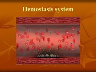
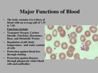
Protective functions of blood Hemostasis system. Hemostatis. Hemostasis or stoppage of bleeding (stasis = halting). No hemostasis No sealing bleed to death from minor wounds The hemostasis response is fast localized and carefully controlled

E N D
Hemostatis Hemostasis or stoppage of bleeding (stasis = halting). No hemostasis No sealing bleed to death from minor wounds The hemostasis response is fast localized and carefully controlled Involves many blood coagulation factors normally present in plasma as well as some substances that are released by platelets and injured tissue cells. During hemostasis, following steps occur: Vascular spasms, Platelet plug formation, Coagulation, or blood clotting. Growth of fibrous tissue in clot to close the hole in vessel. Blood loss at the site is permanently prevented when fibrous tissue grows into the clot and seals the hole in the blood vessel. 2
Coagulation or blood clotting Complicated process, Liquid Blood becomes gel, Over 50 Substances are involved Factors that enhance clot formation are called clotting factors or procoagulants. Factors that inhibit clotting are called anticoagulants. Balance between these two groups of factors. Normally, anticoagulants dominate and clotting is prevented; but when a vessel is ruptured, procoagulant activity in that area increases dramatically and clot formation begins. The procoagulants are numbered I to XIII according to the order of their discovery; hence the numerical order does not reflect the reaction sequence. Most of these factors are plasma proteins made by the liver that circulate in an inactive form in blood until mobilized. Coagulation 5
Three Phases of Coagulation: A complex substance called prothrombin activator is formed. Prothrombin activator converts prothrombin (a plasma protein) into thrombin, (an enzyme). Thrombin catalyzes the joining of fibrinogen molecules present in plasma to a fibrin mesh. Role of Vitamin K in coagulation. Vitamin K not directly involved in coagulation, this fat-soluble vitamin is required for the synthesis of four of the procoagulants made by the liver i.e (II, VII, IX and X). 3-Coagulation 7
Clotting may be initiated by either the Intrinsic Pathway Extrinsic pathway Both pathways are usually triggered by the tissue-damaging events. Clotting of blood outside the body (such as in a test tube) is initiated only by the intrinsic mechanism. Critical components in both mechanisms are negatively charged membranes, particularly those on platelets that contain phosphatidylserine (platelets phospholipids), also known as PF3(platelet factor 3). Many intermediates of both pathways can be activated only in the presence of PF3. Phase 1- Formation of prothrombin Activator 9
Intrinsic Pathway In the slower intrinsic pathway, all factors needed for clotting are present in (intrinsic to) the blood. Extrinsic Pathway By contrast, when blood is exposed to an additional factor in tissues underneath the damaged endothelium called tissue factor (TF), factor III, or tissue thromboplastin, the “shortcut” extrinsic mechanism, which bypasses several steps of the intrinsic pathway, is triggered. Role of calcium Each pathway requires ionic calcium and involves the activation of a series of procoagulants, each functioning as an enzyme to activate the next procoagulant in the sequence. The intermediate steps of each pathway cascade toward a common intermediate, factor X. Activated factor X complexes with calcium ions, PF3, and factor V to form prothrombin activator. Slowest step of the blood clotting process, but once formed, the clot forms in 10 to 15 seconds. Phase 1- Formation of prothrombin Activator 10
Intrinsic Pathway Blood Trauma or contact with collagen XII (Hageman) Activated XII (XIIa) (1) HMW Kininogen, Prekellikerein XI (PTA) Activated XI (XIa) (2) Ca++ IX (PTC) ActivatedIX (IXa) (3) VIII (AHF-A) Thrombin VIIIa (4) Ca++ ActivatedX (Xa) X (SPF) Ca++ Thrombin V (5) Va or PF3 Prothrombin Activator Ca++ Thrombin Prothrombin 11
Extrinsic Pathway Tissue trauma (1) VII (Proconvertin) Activated VII (VIIa) (2) Ca++ (3) X (SPF) ActivatedX (Xa) Ca++ Thrombin V Va Prothrombin Activator Ca++ or PF3 Prothrombin Thrombin 12
Phase 2: Common Pathway to Thrombin Prothrombin activator catalyzes the transformation of the plasma protein prothrombin to the active enzyme thrombin. Intrinsic pathway Extrinsic pathway Prothrombin Activator complex Prothrombin Thrombin 13
Phase 3: Common Pathway to the Fibrin Mesh Thrombin catalyzes the polymerization of fibrinogen (another plasma protein made by the liver). Thrombin is a protein enzyme with weak proteolytic capabilities. It acts on fibrinogen to remove four low-molecular weight peptides from each molecule of fibrinogen, forming one molecule of fibrin monomer. Fibrin monomers has the automatic capability to polymerize with other fibrin monomer molecules to form fibrin fibers. Many fibrin monomer molecules polymerize within seconds into long fibrin fibers. 14
During early polymerization, fibrin fibers are held together by weak non covalent hydrogen bonding, No cross-linkage with one another. fibrin-stabilizing factor causes the cross linkage of fibrin fibers (Released from platelets entrapped in the clot). Activated by thrombin This activated substance operates as an enzyme to form covalent bonds between fibrin monomer molecules, as well as multiple cross linkages between adjacent fibrin fibers. 15
Within 30 to 60 minutes, the clot is stabilized further by a platelet-induced process called clot retraction. Platelets contain contractile proteins (actin and myosin), and they contract in much the same manner as muscle cells. As the platelets contract, they pull on the surrounding fibrin strands, squeezing serum (plasma minus the clotting proteins) from the mass, compacting the clot and drawing the ruptured edges of the blood vessel more closely together. Even as clot retraction is occurring, vessel healing is taking place. Platelet-derived growth factor (PDGF) released by platelet degranulation stimulates smooth muscle cells and fibroblasts to divide and rebuild the wall. As fibroblasts form a connective tissue patch in the injured area, endothelial cells, stimulated by vascular endothelial growth factor (VEGF), multiply and restore the endothelial lining. 4-Clot Retraction and Repair 16
A process called fibrinolysis removes unneeded clots when healing has occurred. Because small clots are formed continually in vessels, this cleanup is important. Without fibrinolysis, blood vessels would gradually become completely blocked. The critical natural “clot buster” is a fibrin-digesting enzyme called plasmin, which is produced when the plasma protein plasminogen is activated. Fibrinolysis 17
Large amounts of plasminogen are incorporated into a forming clot, where it remains inactive until appropriate signals reach it. The presence of a clot in and around the blood vessel causes the endothelial cells to secrete • tissue plasminogen activator (tPA). Along with that • Activated factor XII and • thrombin released during clotting also serve as plasminogen activators. As a result, most plasmin activity is confined to the clot, and any plasmin that strays into the plasma is quickly destroyed by circulating enzymes. • Fibrinolysis begins within two days and continues slowly over several days until the clot is finally dissolved.
Normally, two homeostatic mechanisms prevent clots from becoming unnecessarily large: swift removal of clotting factors, and inhibition of activated clotting factors. Limiting the Activity of Thrombin As a clot forms, almost all of the thrombin produced is bound onto the fibrin threads. This is an important safeguard because thrombin also exerts positive feedback effects on the coagulation process prior to the common pathway. It speed up the production of prothrombin activator by acting through factor V, It also accelerates the earliest steps of the intrinsic pathway by activating platelets. Thus, fibrin effectively acts as an anticoagulant to prevent enlargement of the clot and prevents thrombin from acting elsewhere. Thrombin not bound to fibrin is quickly inactivated by antithrombin III, a protein present in plasma. It inactivates the protease activity of thrombin and factors IXa, Xa, XIa and XIIa by forming complexes with them. Heparin, the natural anticoagulant contained in basophil and mast cell granules, inhibits thrombin by enhancing the activity of antithrombin III. Factors Limiting Normal Clot Growth 19