Download
1 / 33
330 likes | 406 Views
Protective functions of Skin by Dr Aijaz.

E N D
Body’s largest organ; covers area of 1.5 to 2.0 m2; accounts for about 15% of the total body weight;• Consist of 2 layers: epidermis (top) stratified, dermis (deep) connective tissue layer;• Below the skin is the hypodermis (another connective tissue layer, it is not a technical part of the skin)
• Most of the skin is 1-2mm thick. although eyelids(0.5mm) and 6mmbetween the shoulder blades. variation is due to the thickness of thedermis;• Classification of the skin is based on the epidermis alone especially the surface layer of dead cells (stratum corneum): -thick: palm, soles, fingers, and toes;hassweat glands but no hair follicles, nor oil glands(sebaceous); -thin: rest of the body; has hair follicles, sebaceous glands and sweat glands;• Hairless (Glabrous) skin has an epidermal layer of about 1.5 mm inthickness and a dermis of about 3 mm• Hairy skin has an epidermal layer of 0.07 mm in thickness and adermis of about 1-2 mm;
EpidermisConsist of a surface comprised of dead cells packed with keratin (a tough protein);• Does not contain blood vessels. but it contains sparse nerve endings for touch and pain (But most sensations are due to the dermis);• Cell types:– Keratinocytes - produce keratin– Melanocytes - produce melanin
Dermis• Composed mainly of collagen, but also contains elasticreticular fibers, blood vessels, sweat glands, sebaceousglands, hair follicles, nail roots, sensory nerve endings andmuscular tissue (facial expressions are due to the skeletalmuscle connection to the dermal collagen fibers to producesmile, frown, eyebrow movement…)• The boundaries of dermis vs epidermis is not strict, it’s likecardboard ridges that merge together. In sensitive areas, thedermis is more extended and pushed to the surface allowingblood vessels and nerve endings to reach closer to thesurface (look at hand, front vs back)• Functions: Pressure detection; metabolism (duplication ofcells)…
Hypodermis• A sub dermal layer of adipose tissue or otherwise called subcutaneous fat; which is made up of loose, fibrous tissue, rich in blood vessels, lymphatic vessels and nerves;• The base of hair follicles and the coiled tubes of sweat glands may also project down into the hypodermis;• This is the layer that pads the body, serves as an energyreservoir and provides thermal insulation, (it isdifferently distributed in females vs males
White ReactionWhen a pointed object is drawn lightly over the skin, the stroke lines become pale (white reaction). The mechanical stimulus apparently initiates contraction of the precapillary sphincters, and blood drains out of the capillaries and small veins. The response appears in about 15 seconds.
Triple ResponseWhen the skin is stroked more firmly with a pointed instrument, instead of the white reaction there is reddening at the site that appears in about 10 seconds (red reaction).This is followed in a few minutes by local swelling and diffuse, mottled reddening around the injury. The initial redness is due to capillary dilation, a direct response of the capillaries to pressure. The swelling (wheal)is local edema due to increased permeability of the capillaries and postcapillaryvenules, with consequent extravasation of fluid. The redness spreading out from the injury (flare)is due to arteriolar dilation. This three-part response, the red reaction, wheal, and flare, is called the triple response and is part of the normal reaction to injury
The flare is absent in locally anesthetized skin and in denervated skin after the sensory nerves have degenerated, but it is present immediately after nerve block or section above the site of the injury. This plus other evidence indicates that it is due to an axon reflex, a response in which impulses initiated in sensory nerves by the injury are relayed antidromically down other branches of the sensory nerve fibers
Reactive HyperemiaA response of the blood vessels that occurs in many organs but is visible in the skin is reactive hyperemia,an increase in the amount of blood in a region when its circulation is reestablished after a period of occlusion. When the blood supply to a limb is occluded, the cutaneous arterioles below the occlusion dilate.When the circulation is reestablished, blood flowing into the dilated vessels makes the skin become fiery red. O2 diffuses a short distance through the skin, and reactive hyperemia is prevented if the circulation of the limb is occluded in an atmosphere of 100% O2. Therefore, the arteriolar dilation is apparently due to a local effect of hypoxia.
Tissue Macrophages in the Skin and Subcutaneous Tissues (Histiocytes).Although the skin is mainly impregnable to infectious agents, this is no longer true when the skin is broken. When infection begins in a subcutaneoustissue and local inflammation ensues, local tissue macrophages can divide in situ and form still more macrophages. Then they perform the usual functions of attacking and destroying the infectious agents,
Wound HealingWhen tissue is damaged, platelets adhere to exposed matrix via integrins that bind to collagen and laminin.Blood coagulation produces thrombin, which promotes platelet aggregation and granule release. The platelet granules generate an inflammatory response. White blood cells are attracted by selectins and bind to integrins on endothelial cells, leading to their extravasation through the blood vessel walls. Cytokines released by the white blood cells and platelets up-regulate integrins on macrophages, which migrate to the area of injury, and on fibroblasts and epithelial cells, which mediate wound healing and scar formation.Plasminaids healing by removing excess fibrin. This aids the migration of keratinocytes into the wound to restore the epithelium under the scab. Collagen proliferates, producing the scar. Wounds gain 20% of their ultimate strength in 3 weeks and later gain more strength, but they never reach more than about 70% of the strength of normal skin.
Skin as Blood ReservoirShock is more profound in patients with elevated temperatures because of the cutaneousvasodilation, and patients in shock should not be warmed to the point that their body temperature rises.This is sometimes a problem because well-meaning laymen have read in first-aid books that "injured patients should be kept warm," and they pile blankets on accident victims who are in shock.
Control of Skin Coloration and Proctection against Radiation.Fish, reptiles, and amphibia change the color of their skin for thermoregulation, camouflage, and behavioral displays. They do this in part by moving black or brown granules into or out of the periphery of pigment cells called melanophores. The granules are made up of melanins, which are synthesized from dopa and dopaquinone. The movement of these granules is controlled by a variety of hormones and neurotransmittersIn mammals, there are no melanophores containing pigment granules that disperse and aggregate, but there are melanocytes, which have multiple processes containing melanin granules. The melanocytes contain melanotropin-1 receptors, one of several types of melanotropin receptors that have been cloned.
Pigment Abnormalities in Humans Albinos have a congenital inability to synthesize melanin. Albinism occurs in humans and many other mammalian species. It can be due to a variety of different genetic defects in the pathways for melanin synthesis. Piebaldism is characterized by patches of skin that lack melanin as a result of congenital defects in the migration of pigment cell precursors from the neural crest during embryonic development. Not only the condition but also the precise pattern of the loss is passed from one generation to the next. Vitiligo is due to a similar patchy loss of melanin, but the loss develops after birth and is progressive.
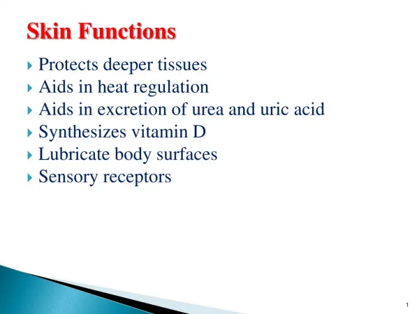
3-WATER PROOFING • In dry environment , it is a mean of preventing fluid loss . • Impermeability of keratin to water is maintained by the thickened lipid wall but some chemicals are lipid soluble & can penetrate . • The secretion of sebaceous glands over the skin & the outer ear by the modified sweat glands or ceruminous glands secrete water repellent wax .
Daily Loss of Body Water Insensible Water Loss. Some of the water losses cannot be precisely regulated. • there is a continuous loss of water by evaporation from the respiratory tract and diffusion through the skin, 700 ml/day of water loss under normal conditions. This is termed insensible water loss because we are not consciously aware of it. • The insensible water loss through the skin occurs independently of sweating and is present even in people who are born without sweat glands; the average water loss by diffusion through the skin is about 300 to 400 ml/day. This loss is minimized by the cholesterol-filled cornified layer of the skin, which provides a barrier against excessive loss by diffusion. • When the cornified layer lost, as occurs with extensive burns, the rate of evaporation can increase as much as 10-fold, to 3 to 5 L/day. For this reason, burn victims must be given large amounts of fluid, usually intravenously, to balance fluid loss.
Vitamin D synthesisThe active transport of Ca2+ and PO43- from the intestine is increased by a metabolite of vitamin D.The term "vitamin D" is used to refer to a group of closely related sterols produced by the action of ultraviolet light on certain provitamins. Vitamin D3, which is also called cholecalciferol,is produced in the skin of mammals from 7-dehydrocholesterol by the action of sunlight.
Insulator System of the BodyThe skin, the subcutaneous tissues, and especially the fat of the subcutaneous tissues act together as a heat insulator for the body. The fat is important because it conducts heat only one third as readily as other tissues.When no blood is flowing from the heated internal organs to the skin, the insulating properties of the normal male body are about equal to three quarters the insulating properties of a usual suit of clothes. Inwomen, this insulation is even better. The insulation beneath the skin is an effective means of maintaining normal internal core temperature, even though it allows the temperature of the skinto approach the temperature of the surroundings.
Blood Flow to the Skin from the Body Core Provides Heat TransferBlood vessels are distributed profusely beneath the skin. Especially important is a continuous venous plexus that is supplied by inflow of blood from the skin capillaries.In the most exposed areas of the body—the hands, feet, and ears—blood is also supplied to the plexus directly from the small arteries through highly muscular arteriovenousanastomoses.The rate of blood flow into the skin venous plexus can vary tremendously—from barely above zero to as great as 30 per cent of the total cardiac output. A high rate of skin flow causes heat to be conducted from the core of the body to the skin with great efficiency, whereas reduction in the rate of skin flow can decrease the heat conduction from the core to very little.
Temperature-Decreasing Mechanisms When the Body Is Too HotThe temperature control system uses three important mechanisms to reduce body heat when the body temperature becomes too great:1. Vasodilation of skin blood vessels. In almost all areas of the body, the skin blood vessels become intensely dilated. This is caused by inhibition of the sympathetic centers in the posterior hypothalamus that cause vasoconstriction. Full vasodilationcan increase the rate of heat transferto the skin as much as eightfold.2. Sweating. The effect of increased body temperature to cause sweating when the body core temperature rises above the critical level of 37°C(98.6°F). An additional 1°C increase in body temperature causes enough sweating to remove 10 times the basal rate of body heat production.
3. Decrease in heat production. The mechanisms thatcauseexcess heat production, such as shivering and chemical thermogenesis, are strongly inhibited.
Temperature-Increasing Mechanisms When the Body Is Too ColdWhen the body is too cold, the temperature control system institutes exactly opposite procedures. They are:1. Skin vasoconstriction throughout the body. This is caused by stimulation of the posterior hypothalamic sympathetic centers.2. Piloerection. Piloerection means hairs “standing on end.” Sympathetic stimulation causes the arrectorpili muscles attached to the hair folliclesto contract, which brings the hairs to an upright stance. This is not important in human beings, but in lower animals, upright projection of the hairs allows them to entrap a thick layer of “insulator air” next to the skin, so that transfer of heat to the surroundings is greatly depressed.
3. Increase in thermogenesis:-(heat production). Heat production by the metabolic systems is increased by promoting shivering, sympathetic excitation of heat production, and thyroxine secretion. These methods of increasing heat require additionalexplanation, which follows.