Download
1 / 29
290 likes | 442 Views
CPR 2000. Dr. THANAPONG HONGPROMYATI. Adult Cardiac Arrest. BLS algorithm if appropriate. Precordial thumb if appropriate. Attach defibrillator/monitor. Assess rhythm. 1. 2. Figure 1. ILCOR Universal/International ACLS Algorithm. Assess rhythm. 3. 4. Check pulse+/-. VF/VT.

E N D
CPR 2000 Dr. THANAPONG HONGPROMYATI
Adult Cardiac Arrest BLS algorithm if appropriate Precordial thumb if appropriate Attach defibrillator/monitor Assess rhythm 1 2 Figure 1. ILCOR Universal/International ACLS Algorithm.
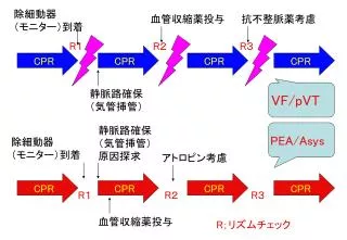
Assess rhythm 3 4 Check pulse+/- VF/VT Non-VF/VT During CPR 5,6 Attempt defibrillation *3 as necessary 3 • Check electrode/paddle position and contact • Attempt to place, confirm, secure airway • Attempt and verify IV access • Patients with VF/VT refractory to initial shocks: - Epinephrine 1 mg IV, every 3-5 min or - Vasopressin 40 U IV, single dose, 1 time only • Patients with non-VF/VT rhythm: - Epinephrine 1 mg IV, every 3-5 min • Consider: buffers, antiarrhythmics, pacing • Search for and correct reversible cause CPR up to 3 min CPR for 1 min Figure 1. ILCOR Universal/International ACLS Algorithm.
Consider causes that are potentially reversible • Hypovolemia • Hypoxia • Hydrogen ion-acidosis • Hyper-/Hypokalemia • Hypothermia • “Tablet” (drug OD,accidents) • Temponade, cardiac • Tension pneumothorax • Thrombosis, coronary (ACS) • Thrombosis, pulmonary (embolism) 7 Figure 1. ILCOR Universal/International ACLS Algorithm.
Person collapses • Possible cardiac arrest • Assess responsiveness Unresponsive Begin Primary ABCD Survey (Begin BLS Algorithm) 1 • Activate emergency response system • Call for defibrillator • A Assess breathing (open airway, look, listen, and feel) No Breathing • B Give 2 slow breaths • C Assess pulse, if no pulse • CStart chest compressions • D Attach monitor/defibrillator when available 1 No pulse Figure 2. Comprehensive ECC Algorithm.
No pulse • CPR continues • Assess rhythm Attempt defibrillation (Up to 3 shock if VF persists) Non-VF/VT (asystole or PEA) 2 3 Secondary ABCD Survey 4,5 • Airway: attempt to place airway device • Breathing: confirm and secure airway device, ventilation, oxygenation • Circulation: gain intravenous access; give adrenergic agent; consider antiarrhythmics, buffer agents, pacing CPR up to 3 min CPR for 1 min Non-VF/VT patients: - Epinephrine 1 mg IV, repeat every 3-5 min VF/VT patients: - Vasopressin 40 U IV, single dose, 1 time only or - Epinephrine 1 mg IV, repeat every 3-5 min • Differential Diagnosis: search for and treat reversible cause Figure 2. Comprehensive ECC Algorithm.
Primary ABCD Survey Focus: basic CPR and defibrillation • Check responsiveness • Activate emergency response system • Call for defibrillator Rhythm after first 3 shocks? 1 A Airway:open the airway B Breathing: provide positive-pressure ventilations C Circulation: give chest compressions D Defibrillation: assess for and shock VF/pulesless VT, up to 3 times (200J,200-300J,360J, or equivalent biphasic) if necessary Figure 3. Ventricular Fibrillation/Pulseless VT Algorithm.
Secondary ABCD Survey A Airway: Place airway device as soon as possible • B Breathing: • Confirm airway device placement by exam plus confirmation device. • Secure airway device; purpose-made tube holders preferred. • Confirm effective oxygenation and ventilation. Focus: more advanced assessments and treatments • C Circulation: • Establish IV access. • Identify rhythm; monitor. • Administer drugs appropriate for rhythm and condition. D Differential Diagnosis: Search for and treat identified reversible causes. Persistent or recurrent VF/VT 2 Figure 3. Ventricular Fibrillation/Pulseless VT Algorithm.
Resume attempts to defibrillate 5 Epinephrine 1 mg IV push, repeat every 3 to 5 minutes or Vasopressin 40 U IV, single dose, 1 time only 3 Resume attempts to defibrillate 1*360J (or equivalent biphasic) within 30 to 60 sec. Consider antiarrhythmics: amiodarone (IIb), lidocaine (Indeterminate), magnesium (IIb if hypomagnesemic state), procainamide (IIb for intermittent/recurrent VF/VT). Consider buffers. 4 Figure 3. Ventricular Fibrillation/Pulseless VT Algorithm.
Antiarrhythmics & Buffer • Amiodarone(class IIb) 300 mg IV push (cardiac arrest dose) If VF/pulseless VT recurs, consider administration of a second dose of 150 mg IV. Max cumulative dose 2.2 g over 24 hr. • Lidocaine (class Indeterminate) 1.0 - 1.5 mg/kg IV push. Consider repeat in 3 to 5 min to a max cumulative dose of 3 mg/kg. • Magnesiumsulfate 1 to 2 g IV in polymorphic VT (torsades de pointes) and suspected hypomagnesemic state. • Procainamide 30 mg/min in refractory VF (Max total dose: 17 mg/kg) is acceptable but not recommended • Sodiumbicarbonate 1 mEq/kg IV is indicated for several conditions known to provoke sudden cardiac arrest.
PULSELESS ELECTRICAL ACTIVITY (PEA = Rhythm on monitor, without detectable pules) Primary ABCD Survey Focus: basic CPR and defibrillation • Check responsiveness • Activate emergency response system • Call for defibrillator A Airway:open the airway B Breathing: provide positive-pressure ventilations C Circulation: give chest compressions D Defibrillation: assess for and shock VF/pulesless VT Figure 4. Pulseless Electrical Activity Algorithm.
A Airway: Place airway device as soon as possible Secondary ABCD Survey • B Breathing: • Confirm airway device placement by exam plus confirmation device. • Secure airway device; purpose-made tube holders preferred. • Confirm effective oxygenation and ventilation. Focus: more advanced assessments and treatments • C Circulation: • Establish IV access. • Identify rhythm; monitor. • Administer drugs appropriate for rhythm and condition. • Assess for occult blood flow (“pseudo-EMT”) D Differential Diagnosis: Search for and treat identified reversible causes. EMD=electro-mechanical dissociation Figure 4. Pulseless Electrical Activity Algorithm.
Review for most frequent causes 1 • Hypovolemia • Hypoxia • Hydrogen ion-acidosis • Hyper-/Hypokalemia • Hypothermia • “Tablet” (drug OD,accidents) • Temponade, cardiac • Tension pneumothorax • Thrombosis, coronary (ACS) • Thrombosis, pulmonary (embolism) Epinephrine 1 mg IV push, repeat every 3 to 5 minutes 2 Atropine 1 mg IV (if PEA rate is slow), repeat every 3-5 minutes as need, to a total dose of 0.04 mg/kg 3 Figure 4. Pulseless Electrical Activity Algorithm.
Primary ABCD Survey Focus: basic CPR and defibrillation • Check responsiveness • Activate emergency response system A Airway:open the airway • Call for defibrillator B Breathing: provide positive-pressure ventilations C Circulation: give chest compressions Confirm true asystole D Defibrillation: assess for VF/pulesless VT; shock if indicate Asystole 1 Rapid scene survey: any evidence personnel should not attempt resuscitation? Figure 5. Asystole: The Silent Heart Algorithm.
A Airway: Place airway device as soon as possible Secondary ABCD Survey • B Breathing: • Confirm airway device placement by exam plus confirmation device. • Secure airway device; purpose-made tube holders preferred. • Confirm effective oxygenation and ventilation. Focus: more advanced assessments and treatments • C Circulation: • Confirm true asystole • Establish IV access. • Identify rhythm; monitor. • Administer drugs appropriate for rhythm and condition. D Differential Diagnosis: Search for and treat identified reversible causes. 2,3 Figure 5. Asystole: The Silent Heart Algorithm.
Transcutaneous pacing If considered, perform immediately 4 Epinephrine 1 mg IV push, repeat every 3 to 5 minutes 5 Atropine 1 mg IV, repeat every 3 to 5 minutes up to a total of 0.04 mg/kg 6 Asystole persists Withhold or cease resuscitation efforts? 7,8,9 • Consider quality of resuscitation? • Atypical clinical features present? • Support for cease-efforts protocols in place? Figure 5. Asystole: The Silent Heart Algorithm.
2 7 • Confirm true asystole- Check lead and cable connection- Monitor power on?- Monitor gain up ?- Verify asystole in another lead • Review the quality of the resuscitation attempt- Was there an adequate trial of BLS? of ACLS? Has the team done the following:- Achieved tracheal intubation?- Performed effective ventilation?- Shocked VF if present?- Obtained IV access?- Given epinephrine IV? Atropine IV?- Ruled out or corrected reversible causes?- Continuously documented asystole >5 to 10 min after all of the above have been accomplished? • Reviewed for atypical clinical features?- Not a victim of drowning or hypothermia?- No reversible therapeutic or illicit drug overdose? 8
Bradycardia • Slow (absolute bradycardia = rate<60bpm • Relatively slow (rate less than expected relative to underlying condition or cause) Primary ABCD Survey • Assess ABCs • Secure airway noninvasively • Ensure monitor/defibrillator is available Secondary ABCD Survey • Assess secondary ABCs (invasive airway management needed?) • Oxygen-IV access-monitor-fluids • Vital sign, pulse oximeter, monitor BP • Obtain and review 12 lead ECG • obtain and review portable Chest x-ray • Problem-focused history • Problem-focused physical examination • Consider cause (differential diagnoses) Figure 6. Bradycardia Algorithm.
Serious sign or symptom? Due to the bradycardia? 1,2 Yes No • Intervention sequence • Atropine 0.5-1.0 mg • Transcutaneous pacing if available • Dopamine 5-20 ug/kg per min • Epinephrine 2-10 ug/min 3,4,5 Type II second-degree AV block or Third-degree AV block? 6 No Yes • Prepare for transvenous pacer • If symptoms develop, use transcutaneous pacemaker until transvenous pacer placed 7 Observe Figure 6. Bradycardia Algorithm.
1 2 • If the patient has serious sign or symptoms, make sure they are related to the slow rate. • Clinical manifestations include- Symptoms (chest pain, shortness of breath, decrease level of consciousness)- Signs (low blood pressure, shock, pulmonary congestion, CHF) • If the patient is symptomatic, do not delay transcutaneous pacing while awaiting IV access or for atropine to take effect • Denervated transplanted hearts will not response to atropine. Go at once pacing, catecholamine infusion, or both. • Never treat the combination of third-degree heart block and ventricular escape beats with lidocaine (or any agent that suppresses ventricular escape rhythms) 3 4 6
Evaluate patient • Is patient stable or unstable? • Are there serious signs or symptoms? • Are signs and symptoms due to tachycardia? Stable Unstable • Unstable patient: serious signs or symptoms • Establish rapid heart rate as cause of signs and symptoms • Rate related signs and symptoms occur at many rates, seldom < 150 bpm • Stable patient: no serious signs and symptoms • Initial assessment identified 1 of 4 type of tachycardia • Atrial fibrillation/flutter • Narrow-complex tachycardia • Stable wide-complex tachycardia: unknown type • Stable monomorphic VT and/or polymorphic VT • Prepare for immediate cardioversion (see algorithm) Figure 7. The Tachycardia Overview Algorithm.
1. Atrial fibrillation Atrial flutter Evaluation focus, 4 clinical features: 1. Patient clinical unstable? 2. Cardiac function impaired? 3. WPW present? 4. Duration<48 or >48 hours? Treatment focus: clinical evaluation 1. Treat unstable patient urgently 2. Control the rate 3. Convert the rhythm 4. Provide anticoagulation Treatment of atrial fibrillation/atrial flutter (See following table) Figure 7. The Tachycardia Overview Algorithm.
2. Narrow-complex tachycardia • Attempt to establish a specific diagnosis • 12 lead ECG • Clinical information • Vagal maneuvers • Adenosine • Diagnosis effort yield • Ectopic atrial tachycardia • Multifocal atrial tachycardia • Paroxysmal supraventricular tachycardia Treatment of SVT (see narrow-complex tachycardia algorithm) Figure 7. The Tachycardia Overview Algorithm.
3. Stable wide-complex tachycardia: unknown type 4. Stable monomorphic VT and/or polymorphic VT • Attempt to establish a specific diagnosis • 12-lead ECG • Esophageal lead • Clinical information Treatment of stable monomorphic and polymorphic VT (see stable VT: monomorphic and polymorphic algorithm) Confirmed SVT Confirmed stable VT Wide-complex tachycardia of unknown type Treatment of SVT (see narrow- complex tachycardia algorithm) Preserved cardiac function Ejection fraction < 40% Clinical CHF Dc cardioversion or Amiodarone Dc cardioversion or Procainamide or Amiodarone Figure 7. The Tachycardia Overview Algorithm.
Control of Rate and Rhythm (Continued From Tachycardia Overview) • AF/flutter with • Normal heart • Impair heart • WPW 2. Control Rhythm 1. Control Rate Duration<48Hrs Duration>48Hrs or Unknown • Consider • DC cardioversion Note: If AF>48 hours’ duration, use agents to convert rhythm with extreme caution in patients not receiving adequate anticoagulation because of possible embolic complications. • NODC cardioversion! • Note: Conversion of AF to NSR with drugs or shock may cause embolization of atrial thrombi unless patient has adequate anticoagulation. • Use antiarrhythmic agents with extreme caution if AF>48 hours’ duration (see note below). or • Use only 1 of the Class IIa following agents (see note below): • Amiodarone • Ibutilide • Flecainide • Propafenone • Procainamide • For additional drugs that are Class IIb recommendation, see Guidelines or ACLS text Normal cardiac function • Use only 1 of the following agents (see note below): • Calcium channel blockers(ClassI) • B-Blockers(ClassI) • For additional drugs that are ClassIIb recommendations, see Guideline or ACLS text • Delayed cardioversion Anticoagulation * 3 weeks at proper levels • Cardioversion, then • Anticoagulation * 4 weeks more or Early cardioversion • Begin IV heparin at once • TEE to exclude atrial clot. then • Cardioversion within 24 h. then • Anticoagulation * 4 more weeks Note:If AF>48hours’ duration, use agents to convert rhythm with extreme caution in patients not receiving adequate anti coagulation because of possible embolic complications. • Consider • DC cardioversion or • Amiodarone (ClassIIb) Impaired heart (EF<40% or CHF) • Anticoagulation as described above, following by • DC cardioversion • Use only 1 of the following agents: • Digoxin (ClassIIb) • Diltiazem (ClassIIb) • Amiodarone(ClassIIb)
Control of Rate and Rhythm (Continued From Tachycardia Overview) • AF/flutter with • Normal heart • Impair heart • WPW 1. Control Rate 2. Control Rhythm Heart Function Preserved Impaired Heart EF<40% or CHF Duration<48Hrs Duration>48Hrs or Unknown Note: If AF>48 hours’ duration, use agents to convert rhythm with extreme caution in patients not receiving adequate anticoagulation because of possible embolic complications. Note: If AF>48 hours’ duration, use agents to convert rhythm with extreme caution in patients not receiving adequate anticoagulation because of possible embolic complications. • Consider • DC cardioversion or • Primary anti- arrhythmic agents Use only 1 of the following agents (see note below**): • Anticoagulation as described above, following by • DC cardioversion WPW • DC cardioversion or • Primary anti- arrhythmic agents Use only 1 of the following agents (see note below): • DC cardioversion or • Amiodarone (ClassIIb) • Amiodarone (ClassIIb) • Flecainide (ClassIIb) • Procainamide (ClassIIb) • Propafenone (ClassIIb) • Sotalol (ClassIIb) • Amiodarone (ClassIIb) • Flecainide (ClassIIb) • Procainamide (ClassIIb) • Propafenone (ClassIIb) • Sotalol (ClassIIb) • Class III (can be harmful) • Adenosine • B-Blockers • Calcium blockers • Digoxin • Class III (can be harmful) • Adenosine • B-Blockers • Calcium blockers • Digoxin
Narrow-Complex Supraventricular Tachycardia, Stable • No DC Cardioversion • Amiodarone • B-Blocker • Ca2+ channel blocker • Attempt therapeutic diagnosis maneuver • Vagal stimulation • Adenosine Preserved EF<40%, CHF • No DC cardioversion • Amiodarone Junctional tachycardia • Priority order: • Ca2+ Channel blocker • B-Blocker • Digoxin • DC cardioversion • Consider procainamide, amiodarone, sotalol Preserved Paroxysmal supraventricular tachycardia • Priority order: • No DC cardioversion • Amiodarone • Diltiazem EF<40%, CHF • No DC cardioversion • Ca2+ channel blocker • B-Blocker • Amiodarone Preserved Ectopic or multifocal atrial tachycardia EF<40%, CHF • No DC cardioversion • Amiodarone • Diltiazem Figure 8. Narrow-Complex Supraventicular Tachycardia Algorithm.
Stable Ventricular Tachycardia Monomorphic or Polymorphic? • Monomorphic VT • Is cardiac function impaired? • Polymorphic VT • Is QT baseline interval prolonged? Note! May go directly to cardioversion Prolong baseline QT interval (suggests torsades) Normal baseline QT interval Normal function Poor ejection fraction • Medications: any one • Procainamide • SotalolOther acceptable • Amiodarone • Lidocaine • Long baseline QT interval • Correct abnormal electrolytesMedications: any one • Magnesium • Overdrive pacing • Isoproterenol • Phenytoin • Lidocaine • Normal baseline QT interval • Treat ischemia • Correct electrolytesMedications: any one • B-Blocker or • Lidocaine or • Amiodarone or • Procainamide or • Sotalol • Amiodarone • 150 mg IV bolus over 10 min. orLidocaine • 0.5 to 0.75 mg/kg IV push. Then use • Synchronized cardioversion Figure 9. Stable Ventricular Tachycardia (Monomorphic or Polymorphic) Algorithm.
Synchronized cardioversion • ventricular tachycardia • Paroxysmal supraventriculartachycardia • Atrial fibrillation • Atrial flutter 100J, 200J 300J, 360J monophasic energy dose (or clinical equivalent biphasic energy dose) Tachycardia with serious signs and symptoms related to the tachycardia If ventricular rate is > 150 bpm, prepare for immediate cardioversion. May give brief trial of medications based on specific arrhythmias. Immediate cardioversion is generally not need if heart rate is <= 150 bpm. • Have available at bedside • oxygen saturation monitor • IV line • Intubation equipment Premedicate whenever possible Figure 10. Synchronized Cardioversion Algorithm.