Download
1 / 16
160 likes | 261 Views
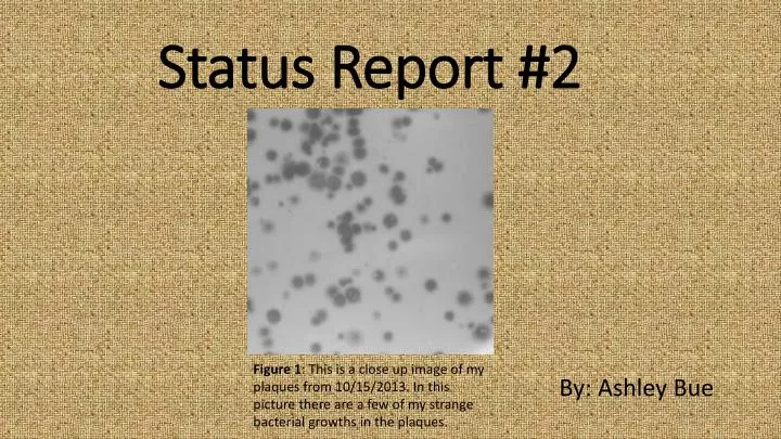
Status Report #2. By: Ashley Bue. Figure 1 : This is a close up image of my plaques from 10/15/2013. In this picture there are a few of my strange bacterial growths in the plaques. Phage Fun Facts. Phages’ play a essential role in the environment and our human lives.

E N D
Status Report #2 By: Ashley Bue Figure 1: This is a close up image of my plaques from 10/15/2013. In this picture there are a few of my strange bacterial growths in the plaques.
Phage Fun Facts • Phages’ play a essential role in the environment and our human lives. • Currently, there are a few selectively studies working to develop current medical conditions. • One of these studies has developed a biodegradable film that is covered with phage. This film has been successful used to cover wounds and prevent bacterial infections. • Also, there is treats using phages being developed for burn victims. Reference: Kirby, Breeann, and Jeremy J. Barr. "Going Viral." The Scientist. www.the-scientist.com, 1 Sept. 2013. Web. 04 Dec. 2013.
Phage Name: Gathje • Named after my High School Biology Teacher, Rochelle Gathje. • She influenced me to pursue a career in biology and through her classes I found my passion for biology. • She even graduated from UWRF! Figure 2: University of Wisconsin-River Falls mascot.
Source of Sample • On 9/8/13 I collected my sample from under a pinetree in a flower bed with cedar mulch. The top soil appeared dry and it contained numerous roots and pieces of mulch. • The weather was about 70⁰ F with an overcast sky but not raining. • GPS Coordinates: 43⁰ 49’ 31 N 91⁰ 58’ 21” W • 10 Minutes outside of Lanesboro, MN Figure 3: Pictured is my hometown of Lanesboro, MN
Development of Plaques Figure 7: 10/15/13 plaques which contain no bacterial growth. Figure 4: These are my first major plaque growths with the bacterial spots. Plaques from 9-26-13 Figure 5: Plaque from 10/3/12 that contains a bacterial growth. Figure 6: Spot test from 10/8/13 all plaques contain the bacterial growth. Figure 8: Plaques from 10/12/13 show growth again after redoing previous procedure because of contamination. Figure 9: Plaques from 10/22/13, one of my final plates.
Plaque Morphology • Titer of HTL: 5.4x10^9 • Size: 8 to 10 mm • Appearance: Clear, Round • Had Strange Appearance to Start Figure 9: This is my 10^ -5 HTL Titration plate created on 10/22/2013.
DNA Isolation • Concentration: 0.1775 μg/μl • Yield: 17.75 μg • A260/280: 1.699 • The ratio of A260/280 should be around 1.8 so mine is a little low but the number is still good. • I have enough DNA to preform all procedures.
Restriction Enzyme Results #1 Gel Electrophoresis #1 1 Kb Ladder Undigested DNA BamHI ClaI EcoRI HaeIII HindIII Figure 10: My first gel results from 10/31/13.
Analysis Fragment Lengths for Restriction Enzymes #1 • When looking at this gel, I can determine that my phage is possible related to three other phages in the class, SJD, JD, and TT. • With only these results it is hard to determine which ones are related or the same phage. • By studying the phages further I should be able to determine which ones are related. Figure 11: This chart depicts my fragment length from my first gel.
Restriction Enzyme Results #2 Gel Electrophoresis #2 PST I SAL I ECO RV NCO I KB Ladder Figure 12: This is my second gel from 11/12/13.
Fragment Length for Restriction Enzymes #2 Figure 13:
Analysis of Second Set of Restriction Enzymes • From the second gel, I am able to conclude that it is not related to SJD and JD. • My gel is completely different from both of these gels, both are picture below. Figure 14: JD gel had different cuts than mine and SJD. Figure 15: SJD gel had different cuts than mine and JD.
Electron Microscope Results • 1, 2 are normal siphoviridae phages. • 3 is missing the DNA inside its head. • 4 has a broken open head which has allowed the dye to enter. • 5 is a normal siphoviridae phage with a curled tail. 2 Phage 1: Tail-96.88nm Head-43.75 nm Phage 2:Tail-100nm Head-40.63nm Phage 3:Tail-100 nm Head-43.75 nm 1 4 5 3 Figure 16: This is my image from the electron microscope.
Final Conclusions • For all of this information I can conclude that my phage is for sure not related to JD or SJD because of the second gel. • A third gel was ran to determine if mine and TT were related. This gel showed very poor results so I can not determine if the two are related. Also there is not a good electron microscope image of TT so I can’t clearly determine if the two images look similar. • My phage shows no exact similarities to any sequenced phage in the database.
Interesting! • My phage started to develop strange bacterial growths in the center of my plaques. • Slowly these growths started to disappear as a purified the phage. Figure 17: This is a streak plate created from the bacterial growths in the center of my plaqes.
WHY MY PHAGE! • Unique bacteria growth! • I have enough DNA. • I have take accurate notes on all procedures. • My phages is not taken from on of Karen’s samples.