Download
1 / 64
2.61k likes | 5.37k Views
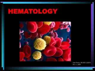
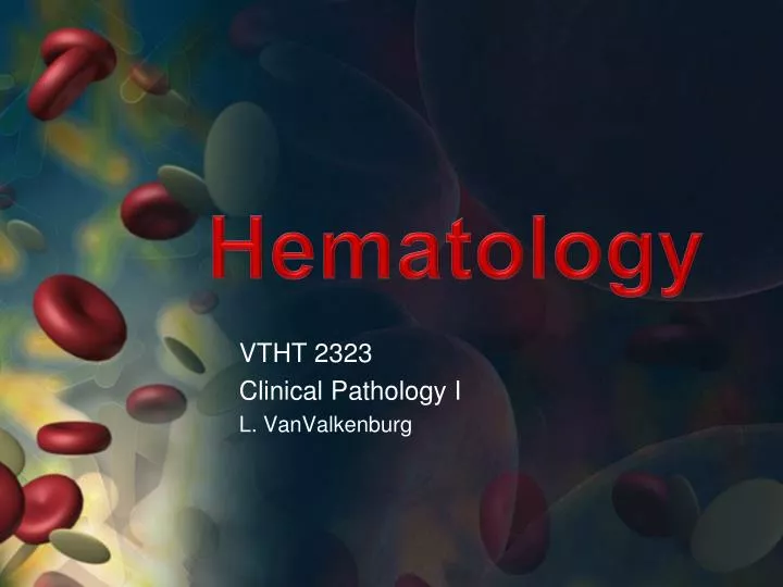
Hematology. VTHT 2323 Clinical Pathology I L. VanValkenburg. General Functions/Purpose of Blood:. Transportation: Dissolved gases, waste products, hormones, enzymes, nutrients, plasma proteins, platelets. etc. Defense: Phagocytosis and other roles in immunity Clotting factors.

E N D
Hematology VTHT 2323 Clinical Pathology I L. VanValkenburg
General Functions/Purpose of Blood: • Transportation: • Dissolved gases, waste products, hormones, enzymes, nutrients, plasma proteins, platelets. etc. • Defense: • Phagocytosis and other roles in immunity • Clotting factors
Regulation: • Maintains body temperature Normal = 101.0 – 102.5 F • Helps to control the body’s pH • Normal pH = 7.35 – 7.45 • Higher pH = alkalosis • Lower pH = acidosis • Maintains homeostasis: a state of chemical equilibrium in the body in response to internal and external changes.
Whole Blood is a viscous substance (fluid) that contains a:1. fluid portion (plasma)2. cellular portion *Depending on the species, plasma makes up 45% - 78% of the total blood volume.
Plasma • Carbohydrates • Vitamins • Hormones* • Enzymes • Lipids • Amino acids* • Salts • Gases • Waste materials • Antibodies* • Other ions and molecules 91% water 7% proteins 2% other dissolved solutes
Cellular Portion of Blood • Erythrocytes • Account for ~45% blood volume (dogs) and ~37% blood volume (cats) • Thrombocytes • 200,000 – 500,000 /µL = dog • 300,000 – 700,000 /µL = cat • Leukocytes • 6,000 – 17,000 /µL = dog • 5,500 – 19,500 /µL = cat
Hematopoiesis • General term for production of all blood cells. • Constant process; blood cells are not immortal. • Mostly occurs in the bone marrow. • Common ancestor of all blood cells is the pluripotent stem cell (PPSC) a.k.a. hematopoietic stem cell. • One PPSC will divide and eventually become many blood cells.
Quiz • What are the three primary functions of blood? • What cells make up the cellular portion of blood? • What is contained in plasma? • What is the term used to describe production of all blood cells? • What is the common ancestor of all blood cells? • Where is blood produced? • Where is blood stored?
Red Blood Cells • Function: • Carry oxygen from the lungs to the tissues • Carry carbon dioxide away from tissues to the lungs and kidneys • The RBC is a vehicle for hemoglobin which is the carrier molecule for oxygen. • The bioconcave shape is a result of the cooperative binding from the 4 heme groups.
Hemoglobin • Every heme group can carry a molecule of oxygen; 4 heme groups attach to each globin molecule. So each hemoglobin molecule can carry 4 molecules of oxygen. • Immature RBCs do not contain their full complement of hemoglobin yet • Rule of Thumb: Hemoglobin accounts for about 1/3 of the RBC (PCV) • Hemoglobin = heme (iron) + globin (protein) • Iron is responsible for color of RBC
Recipe for Red Blood Cells • Water • Iron (for the synthesis of heme~) • Copper (important in release of iron from tissues into plasma) • Protein/amino acids (~globin) • Essential Fatty Acids • Vitamin B6 • Vitamin B12 • Folic Acid (B9)
Erythropoiesis • Formation of erythrocytes. • Occurs in the bone marrow of normal adult animals. • Occurs in the spleen and liver of the fetus. • Maturation time usually takes 5 days. • Average lifespan of RBC is about 110 days for dogs and about 68 days in the cat. • Erythropoiesis is a constant process.
Erythropoiesis 1. Pluripotent stem cells (PPSC) exist in bone marrow 2. Erythropoietin (EPO) (a hormone) is released by kidneys in response to blood hypoxia (kidney dysfunction can affect RBC production) 3. PPSC stimulated by EPO will begin to undergo mitiotic division and develop into myeloid stem cells.
Erythrocyte Maturation • Rubriblast– (14-19 µm) First morphologically identifiable RBC precursor • Prorubricyte - (12-17 µm) • Rubricyte - (12-15 µm) • Metarubricyte - (8-12 µm) - a.k.a. NRBC (nucleated red blood cell) - Released in cases of severe anemia • Reticulocyte - (7-10 µm) • Not nucleated; contains nuclear remnants (Residual RNA) • A.k.a. – polychromatophil (no NMB) • Mature RBC- (7-8 µm)
Canine Bone Marrow Aspirate Bone marrow aspirate demonstrating erythropoiesis. Rubriblast (arrow), prorubricytes (arrowhead), and various stages of rubricytes (nucleated erythroid precursors) are present.
Lifespan and Destruction of RBCs • Lifespan of RBC = related to metabolism and weight of animal • Process of aging is called senescence. • As RBC becomes more senescent, enzyme activity decreases • Glycolytic enzyme unable to break down glucose • Cell becomes more round • Volume decreases • 90% of destruction of senescent RBCs occurs by extravascularhemolysis.
Normal Removal of Aged RBCs • Surface membrane alterations (oxidative damage) or damaged cells are recognized by macrophages. • Macrophages found all over the body (mostly of the spleen) phagocytize (engulf and “eat up”) old RBCs. • Components of RBC breakdown are either removed by the body as waste or recycled.
ExtravascularHemolysis • Hemoglobin is disassembled within minutes after phagocytosis begins • Iron is made available for re-use by the bone marrow • Amino acids return to the protein pool of the body • Carbon is released through lungs as carbon monoxide • Bilirubin is bound to albumin and transported to liver for conjugation.
Bilirubin • In the liver, bilirubin is conjugated – joined to glucuronic acid -- and becomes water soluble. • Conjugated bilirubin is excreted as a bile pigment into the intestines. • In the intestines, it is converted into urobilinogen by bacteria. • Some urobilinogen is reabsorbed by kidneys and eliminated in the urine. • Some urobilinogen is converted to stercobilinogen and excreted in the stool. • These compounds are responsible for the color of urine and feces.
Intravascular Hemolysis • 10% of normal RBC destruction takes place by intravasuclarhemolysis. • Haptoglobinpicks up hemoblobin and carries it to the mononuclear phagocyte system in the liver for further breakdown. • Hemoglobinemia = When all haptoglobin is filled with hemoglobin, excess hemoglobin appears in the plasma • Hemoglobiniuria = Hemoglobin cannot get to the liver so it is carried to the kidney and eliminated in the urine.
Other Causes of Intravascular Hemolysis • Immune Mediated Hemolytic Anemia (IMHA) • RBC parasites (i.e. Babesia sp.) • Some bacteria (i.e. Clostridium, Leptospira) • Bee stings, snake envenomation • Some toxins (i.e. zinc toxicity in dogs) • Acute liver disease • Insulin therapy with some diabetic cats • Congenital defects ** Note that red cells can also lyse or rupture in vitro (either in the blood collection tube or during collection). When this occurs, the hemolysis is considered artifactualhemolysis.
Jaundice/Icterus • Excessive RBC breakdown = hyperbilirubinemia (excess unconjugatedbilirubin in plasma). • If there is too much bilirubin in the blood for the liver to be able to break down, it is deposited in the tissues. • Bilirubin in the tissues = jaundice; clinically seen as a yellowish color in the MM and sclera of the eyes • In liver disease, the liver is unable to break down a normal amount of bilirubin from normal RBC destruction and jaundice results.
Anemia – Not enough RBCs • Pathological condition that results in decreased oxygen-carrying capacity of the blood. • Regenerative • Caused by blood loss or hemolysis • Non-regenerative • Caused by reduced or defective erythropoiesis • Defective hemoglobin production
Polycythemia– More RBCs than needed • An increase above normal in the number of RBCs • Three types: • Relative • Hemoconcentration – loss of fluid from blood • Compensatory • Resulting from chronic hypoxia • Ex: High altitudes, CHF • Polycythemiarubravera • Rare bone marrow disorder characterized by increased production of RBCs for unknown reason.
Quiz • What is the recipe for RBCs? • What stimulates the production of RBCs? • As an RBC matures, what happens to its size? Color? Nucleus? What else? • What happens to an RBC as it becomes senescent? • Where does bilirubin come from? How is it eliminated from the body? • What causes jaundice/icterus? • What are some causes of intravascular hemolysis? • What are some causes of anemia? • What are the three different types of polycythemia?
Erythrocyte Morphology • Different species have differentsized RBCs with varying degrees of central pallor. • Dogs have largest RBCs (~7 µm in diameter) • Cats, horses, cows, sheep, goats (3-4 µm) • Llamas and camels have elliptical (oval) RBCs • Deer have sickle-shaped RBCs • Birds, fish, amphibians, and reptiles have nucleated, elliptic RBCs. • Human RBCs are about the same size as those of dogs.
Normal RBC Morphology • Normocytes- cells look normal • Normochromasia- suggests adequate amount of hemoglobin and typical MCHC Canine blood, normal erythrocyte morphology. Canine RBCs are all about the same size, shape, and color, and have a prominent area of central pallor
Normal RBC Morphology Feline blood. Normal erythrocyte morphology. Feline RBCs are smaller than dog erythrocytes, exhibit a slight amount of crenation, and have a minimal area of central pallor
RBC Morphology • Morphologic characteristics of RBCs can be categorized according to: • Cell arrangement on blood film • Size • Color • Shape • Presence of structures in or on the RBCs
Rouleaux Formation • Grouping of RBCs in “stacks” that resemble coins • Marked rouleaux formation is seen in healthy horses. • Seen with increased fibrinogen or globulin concentrations • May be seen as an artifact in blood that has been held too long before preparing the blood film and in blood that has been refrigerated.
Agglutination • Occurs in immune-mediated disorders • Antibody coats the erythrocyte, resulting in bridging and clumping of RBCs • To differentiate between rouleaux and agglutination, add a drop of saline to a drop of blood and examine microscopically for agglutination. • Rouleaux will disperse in saline.
Anisocytosis • Variation in the size of RBCS • May indicate presence of macrocytes, microcytes, or both. • Common finding in normal bovine blood.
Macrocytosis • Erythrocytes are larger than normalwith an increased MCV. • Usually immature, polychromatophilic RBCS (reticulocytes) • Seen in the presence of regenerative anemia. • Typically polychromasia is also present
Microcytosis • Microcytes are RBCs with a diameter less than that of normal erythrocytes, with a decreased MCV. • Usually occurs with iron deficiency • Normal finding in shibainu and akita dogs
Polychromasia • Recall that the younger the cell in the RBC maturation series, the more dark blue the staining due to increased metabolic activity. • Mature RBCs stain red because they have their full complement of hemoglobin. • Hb production begins right before cell loses nucleus; immature RBCs with some Hb present stain purple/lavender because there is still some metabolic activity going on within the cell.
Hypochromasia • RBCs that have decreased staining intensity caused by insufficient hemoglobin within the cell. • Cell will normally appear more darkly stained along the periphery, gradually tapering to a much paler central region. • Frequently observed in iron deficiency anemia caused by chronic blood loss or parasitism. • Animals with hypochromasia almost always have microcytosis as determined by a decreased MCV.
Hyperchromasia • Refers to cells that appear more darkly stained than normal RBCs. • Gives appearance of oversaturation with Hb and suggests an increase in MCHC. • Because RBC has a fixed maximum capacity for Hb, oversaturation cannot occur. • Presence of hemolysis, Heinz bodies, and lipemia can interfere with tests and artifactually increase MCHC • Spherocytes (arrow below) have the illusion of being hyperchromic and microcytic but still have a normal cell volume
Spherocytes • Dark red staining, appear smaller than average sized RBC, round, lacks central pallor • Not usually detected in species other than dogs. • Common in immune-mediated hemolytic anemia (IMHA) • Antibodies recognize “self” as foreign and attach themselves to surface of RBCs • May also be seen after transfusion with mismatched blood
Canine blood. Regenerative anemia with spherocytes. Anisocytosis is due to macrocytic cells and spherocytes, which are smaller than normal and lack central pallor. Spherocytes are associated with hemolytic anemias due to immune disease or fragmentation. The polychromatophilic RBC with a rod- shaped area of central pallor (arrow) is a stomatocyte
Poikilocytosis • Variations in the shapes of RBCs (poikil ~ irregular) • A general term to encompass nondescript variations in shapes of erythrocytes that are scattered throughout the blood film. • Term does not suggest a specific diagnosis. Note: poikilocytosis, anisocytosis, and polychromasia are present on this slide
Schistocytes • RBC fragments resulting from shearing of red blood cell by intravascular trauma. • May be seen with Disseminated Intravascular Coagulopathy (DIC) or with iron deficiency • Expect to also see thrombocytopenia on blood film of animals with DIC Canine blood. Schistocytes. Fragmentation of RBCs results in the formation of schistocytes. These cells are formed when RBCs are sheared by convoluted vascular channels, intravascular fibrin deposition, or excessive turbulence. The RBCs have sharp cuts in the cell margin that produce membrane tags and cells that have a helmet shape
Acanthocytes(“Spur Cells”) have irregular membrane projections of variable length and diameter with rounded tips. Unevenly sized and spacedMay be seen in blood smear of Cats with hepatic lipidosis, Dogs with liver diseaseor hemangiosarcoma of the liverRepresent in vivo alteration rather than artifact. Canine blood. acanthocytes (arrows). A few spherocytes are also noted.