Download
1 / 1
10 likes | 159 Views
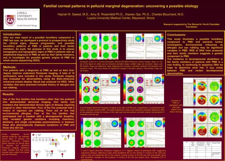
F amilial corneal patterns in pellucid m arginal degeneration: uncovering a possible etiology Hajirah N. Saeed , M.D., Amy B. Rosenfeld Ph.D., Xiaowu Gai , Ph.D., Charles Bouchard, M.D. Loyola University Medical Center , Maywood, Illinois .

E N D
Familial corneal patterns in pellucid marginal degeneration: uncovering a possible etiology Hajirah N. Saeed, M.D., Amy B. Rosenfeld Ph.D., XiaowuGai, Ph.D., Charles Bouchard, M.D. Loyola University Medical Center, Maywood, Illinois Research supported by The Richard A. Perritt Charitable Foundation Introduction: After our case report of a possible hereditary component in PMD last year, we developed a protocol to prospectively study the clinical signs, disease progression, and possible hereditary patterns of PMD in patients and their family members. As such, the purpose of this study is to assess tomographic and topographic signs of PMD in patients with an established diagnosis of PMD as well as their family members, and to investigate the possible genetic origins of PMD via whole exomesequencing (WES). Conclusions: This study illustrates a possible hereditary component to PMD and warrants further investigation. Environmental influences as allergies and eye rubbing may be significant variables in PMD. WES of DNA from probands and their family members suggests a genetic component to PMD. The incidence of developmental disabilities in the family members of patients with PMD is a new finding. In continuing to sequence DNA we hope to determine what link, if any, exists between PMD and certain developmental abnormalities. Pentacam scans of a normal participant utilizing the Belin-Ambrosioectasia display. Maps on the top of the figure for either eye are baseline elevation maps, middle maps are exclusion maps, and the bottom maps are difference maps depicting the change in elevation between the baseline and exclusion maps. This scan shows no significant change in elevation (green). Pachymetric maps are not shown. Methods: Five patients with a diagnosis of PMD as well as their first-degree relatives underwent Pentacam imaging. A total of 16 participants were included in this study. Pentacam imaging was evaluated for abnormalities using the Belin-Ambrosio enhanced ectasia display. Blood was analyzed via WES. Other variables that were assessed included history of allergies and eye rubbing. Results: Two of the five families had members other than the proband who demonstrated abnormal imaging. One family had members that demonstrated clinical signs of disease requiring surgical or other intervention. Four of the five probands had a history of vigorous eye rubbing. Three out of five had environmental allergies. Four of the five families that participated had a member with a developmental disability. WES revealed genetic variations including insertions, deletions, and single nucleotides polymorphisms in between those who phenotypically displayed symptoms of PMD and those who did not. Example of PMD on topography with crab claw pattern and inferior thinning A and B depict abnormal Pentacam imaging in the right and left eyes, respectively, in a participant with known, clinically manifest PMD. The bottom difference maps show significant elevation difference (ED) on the posterior corneal (K) surface of the right eye (red and yellow) and both anterior and posterior K surfaces in the left eye. Family pedigrees. Arrows indicate probands; red indicates clinical signs of disease and abnormal Pentacam imaging; yellow indicates no clinical signs of disease but abnormal Pentacam imaging. Questions marks indicate that that family member was unable to be tested with the Pentacam. C and D depict abnormal Pentacam imaging in a 1st degree relative of participant A/B, suggesting subclinical disease. Difference maps show borderline ED on the anterior K surface of the right eye (yellow only) and significant ED on the anterior corneal surface of the left eye (red and yellow) along with borderline changes on the posterior K surface of the left eye (yellow only). Participant C/D is asymptomatic.