Download
1 / 97
1.11k likes | 1.66k Views
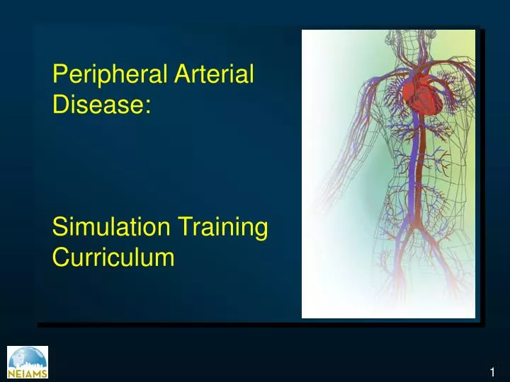
Peripheral Arterial Disease: Simulation Training Curriculum. Peripheral Arterial Disease. Etiology Epidemiology Physical examination Clinical Manifestations Diagnosis Indications Treatment Options Prognosis. Peripheral Arterial Disease: Etiology.

E N D
Peripheral Arterial Disease: Simulation Training Curriculum
Peripheral Arterial Disease Etiology Epidemiology Physical examination Clinical Manifestations Diagnosis Indications Treatment Options Prognosis
Peripheral Arterial Disease: Etiology • Atherosclerosis • Degenerative diseases: Marfan and Ehlers-Danlos syndrome, neurofibromatosis, arteriomegaly • Dysplastic disorders: Fibromuscular dysplasia • Vascular inflamation : Takayasu’s disease • In situ thrombosis • Thromboembolism Hirsh et al Circulation 2006; 113(11): e463-654 ACC/AHA Guidelines
Atherosclerosis: A Progressive and Systemic Process PlaqueRupture/Fissure &Thrombosis Occlusive AtheroscleroticPlaque Unstable Angina FattyStreak FibrousPlaque Normal MI Coronary Death Stroke Effort Angina Claudication Renovascular Dz Clinically Silent Critical Leg Ischemia Increasing Age
Peripheral Arterial Disease: Overlap of Atherosclerotic Disease 38% overlap > 2 Vascular beds Patients with one manifestation often have coexisted disease in other vascular beds Ness et al Am J Geriatr Soc 1999; 47:1255-6
SF-36 Physical Function Scores IntermittentClaudication Average Adult Average Well Adult CLI CHF COPD No. of People 30 50 34 36 38 55 Physical Component Score Adapted from Ware JE. Ann Rev Pub Health. 1995;16:327-354.
Atherosclerosis Disease in the U.S. Ness J et al J Am Geriat Soc 1999; 47: 1255-1256
Risk of Ischemic Events in Atherosclerotic Clinical Syndromes *Sudden death defined as death documented within 1 hour and attributed to Coronary Heart Disease (CHD) † Includes only fatal MI and other CHD; does not include nonfatal MI 1. Adult treatment Panel II Circulation 1994; 89: 1333 – 1435 2. Kannel et al J Cardiovasc Risk 1994;1:333-339 3. Witerdink et al Arch Neurol 1992; 49: 857 – 863 4. Criqui et al N E J Med 1992; 326: 381-386
Peripheral Arterial Disease Etiology Epidemiology Physical examination Clinical Manifestations Diagnosis Indications Treatment Options Prognosis
Prevalence of PAD in 1990s Projected US Age Abnormal ABI Prevalence 40-59 3% 2.1 million 60-69 8% 1.6 million >70 19% 4.7 million Total 8.4 million Criqui et al N Engl J Med 1992;326:381-86 Hiatt et al Circulation 1995;91:1472-79
Peripheral Arterial Disease Prevalence by age PAD Prevalence Age Groups Criqui et al Circulation 1985; 71: 510-5 (77)
Reduced Increased Risk Factors for PAD Smoking Diabetes Hypertension Hypercholesterolemia Hyperhomocysteinemia Fibrinogen C-Reactive Protein Alcohol Relative Risk .5 1 2 3 4 5 6 Newman et al Circulation 1993;88:837-845. Hiatt WR et al Circulation 1995;92:614-621. Graham et al JAMA 1997;277:1775-1781. TASC Working Group J Vasc Surg 2000;31(1, pt 2):S1-S288. Ridker PM et al Circulation 1998;97:425-428.
Nobel Risk Factors as Predictors of PDA Adjusted for age, smokin, DM, family history, HTN, exercise level, and BMI Ridker et al JAMA 2001; 285:2481-5 Pradhan et al Circulation 2002; 106: 820-5
Lower Extremity PAD: Prevalence • Affects a large proportion of most adult populations worldwide • Increases with age and with exposure to atherosclerotic risk factors. • Defined by • Claudication as a symptomatic marker • Abnormal ankle-to brachial systolic blood pressure • Underlying atherosclerosis risk factor profile • Presence of other concomitant manifestations of atherosclerosis Hirsh et al Circulation 2006; 113(11): e463-654 ACC/AHA Guidelines
Risk of developing lower extremity PAD Hirsh et al Circulation 2006; 113(11): e463-654 ACC/AHA Guidelines
Prevalence of intermittent claudication in various studies Dormandy JA, Rutherford RB J VAsc Surg 2000;31: S1-S296
Mean Prevalence of intermittent claudication in large population studies Age group Dormandy JA, Rutherfors RB J Vasc Surg 2000; 31: S1-S296
Peripheral Arterial Disease Etiology Epidemiology Physical examination Clinical Manifestations Diagnosis Indications Treatment Options Prognosis
CVD CAD RVD PAD
Peripheral Arterial Disease: Clinical Diagnosis • Must have a high index of suspicion • Must perform a thorough physicial examination • Determine the global atherosclerotic burden • Utilize the vascular diagnostic laboratory • Magnetic resonance angiography is rapidly replacing invasive testing • Reserve arteriography for cases requiring intervention TCT 2005
Vascular Review of Systems • Any exertional limitation of the lower extremity muscles or any history of walking impairment (fatigue, numbness, aching, or pain • Any poorly healing or non healing of the legs or feet • Any pain at rest localized at the lower leg or foot and its association with the upright or recumbent positions • Postprandial abdominal pain that reproducibly is provoked by eating and is associated with weight loss • Family history of a first-degree relative with Abdominal Aortic Aneurysm ACC/AHA Guidelines
Vascular Physical Examination • Measurement of blood pressure in both arm and notation of any interarm assymetry • Palpation of the carotid pulses and notation of the carotid upstroke and amplitude and presence of bruits • Auscultation of the abdomen and flank for bruits • Palpation of the pulses at the brachial, radial ulnar, femoral, popliteal, dorsalis pedis, and posterior tibial sites. Performance of Allen’s test when knowledge of hand perfusion is needed • Auscultation of both femoral ateries for the presence of bruits • Pulse intensity should be recorded numerically: 0, absent; 1, diminished; 2, normal; 3, bounding • Additional findings: distal hair loss, trophic sin changes hypertrophic nails ACC/AHA Guidelines
Physical Examination Dorsalis Pedis Popliteal Artery Femoral Pulse Posterior Tibial Beard JD. BMJ. 2000;320:854.
Signs of PAD • Decreased or absent pulses • Bruits • Muscle atrophy • Pallor of feet with elevation • Dependent rubor • Signs of chronic ischemia: Hair loss, thickened nails, smooth & shiny skin, coolness, pallor or cyanosis
Peripheral Arterial Disease Etiology Epidemiology Physical examination Clinical Manifestations Diagnosis Indications Treatment Options Prognosis
Clinical Presentation of PAD Patients Chronic limb ischemia Acute Limb Ischemia Asymptomatic PAD Stable Claudication Hirsch AT. Fam Pract Recertification. 2000;15(suppl):6-12.
Lower Extremity Arterial Disease in the Population > 55 y Weitz et al Circulation 1996; 94(11):3026-3049
Individuals at Risk for Lower-extremity Peripheral Arterial Disease • Age less than 50 years, with diabetes and one other atherosclerosis risk factor (smoking, dyslipidemia, hypertension, or hyperhomocysteinemia) • Age 50 to 69 years and history of smoking or diabetes • Age 70 years and older • Leg symptoms with exertion (suggestive of claudication) or ischemic rest pain • Abnormal lower extremity pulse examination • Known atherosclerotic coronary, carotid, or renal artery disease ACC/AHA Guidelines
Common Sites of Claudication 25-30% 80-90% Tibial and peroneal arteries 40-50% Foot Adapted from TCT 2005
Classification of Peripheral Arterial Disease RUTHERFORD Dormandy JA, Rutherfors RB J Vasc Surg 2000; 31(1): S1-S296
Pathophysiology of Intermittent Claudication Intermittent claudication is associated with: • Metabolic abnormalities stemming from reduced blood flow and O2 delivery1 • Significant reduction (50%) in muscle fibers compared with controls2 • Smaller type I and II muscle fibers with greater arterial ischemia2 • Hyperplastic mitochondria and demyelination of nerve fibers3 1 Lundgren et al Am J Physiol. 1988;255:H1156-64. 2 Hedberg et al. Eur Vasc Surg. 1989;3:315-22. 3 Farinon et al. Clin Neuropathol 1984;3:240-52.
PAD Symptom Severity • Maximal walking speed • Normal = 3-4 mph • PAD = 1-2 mph • Maximal walking distance • Normal = unlimited • PAD, 31% difficulty walking in home • PAD, 66% difficulty walking 1/2 block • Peak VO2 • PAD reduced 50% (NYHA class III CHF) Otsuka data set, J Appl Physiol 1992;73:346
Peripheral Arterial Disease Etiology Epidemiology Physical examination Clinical Manifestations Diagnosis Indications Treatment Options Prognosis
Diagnostic Methods • Ankle-and Toes – Brachial Indices, segmental pressure examination • Pulse volume recording • Continuous wave doppler ultrasound • Treadmill exercise testing with and without ABI assessments and 6 minute walk test • Duplex ultrasound • Computed tomographic angiography • Magnetic resonance angiography • Contrast angiography Hirsh et al Circulation 2006; 113(11): e463-654 ACC/AHA Guidelines
Typical Noninvasive Vascular Laboratory Tests for Lower Extremity PAD Patients by Clinical Presentation ACC/AHA Guidelines Adapted from primary cardiology, 2nd ed., Braunwald E, Goldman L, eds. “Recognition and management of peripheral arterial disease”.
Diagnostic Algorithm for PDA History, Physical examination Suggestive of PDA? Search for alternate diagnosis NO Yes Ankle-Brachial Index >0.9 >1.30 <0.9 • Vascular Lab Referral • Segmental pressures, PVR • Graded treadmill test Still suspicious? Anatomic Assessment: DUS, MRA, CTA PAD TCT 2005
Diagnosis of asymptomatic PAD and Atypical Leg Pain Evaluate other causes of leg symptoms Confirmation of PAD diagnosis ACC/AHA Guidelines
Diagnosis of Claudication Classic claudication symptoms: Muscle fatigue, cramping, or pain that reproducibly begins during exercise and that promptly resolves with rest Chart document the history of walking impairment and specific lifestyle limitations Document pulse examination Exercise ABI (TBI, segmental pressure, or duplex ultrasound examination) ABI >0.90 ABI ABI less than or equal to 0.90 Abnormal results Normal results No PAD or consider arterial entrapment syndromes Confirmed PAD diagnosis ACC/AHA Guidelines
Diagnosis of Acute Limb Ischemia Rapid or sudden decrease in limb perfusion threatens tissue viability History and physical examination; determine time of onset of symptoms Emergent assessment of severity of ischemia: Loss of pulses Loss of motor and sensory function Vascular laboratory assessment ABI, TBI, or duplex ultrasound • Severe PAD documented: • ABI less than 0.4 • Flat PVR waveform • Absent pedal flow Nor or minimal PAD Consider and evaluate source of: atheroembolism, thromboembolism or phlegmasia cerulea dolens ACC/AHA Guidelines
Diagnosis of Critical Limb Ischemia Chronic Symptoms: Ischemic rest pain, gangrene, nonhealing wound Ischemic etiology must be established promptly: By examination and objective vascular studies Implication: Impending limb loss • History and physical examination: • Document lower extremity pulses • Document presence of ulcers or infection Assess factor that may contribute to limb risk: diabetes, neuropathy, chronic renal failure, infection ABI, TBI, or duplex ultrasound Severe lower extremity PAD ABI less than 0.4; flat PVR waveform; absent pedal flow No or minimal atherosclerotic arterial occlusive disease ACC/AHA Guidelines
The Ankle-Brachial Index • The resting ABI should be used to stablish the lower extremity PAD diagnosis in patients with suspected lower extremity PAD • Exertional leg symptoms • Non healing wounds • 70 years and older or 50 years and older with history of smoking or diabetes • ABI should be measured in both legs in all new patients with PAD of any severity to confirm the diagnosis and establish a baseline Hirsh et al Circulation 2006; 113(11): e463-654 ACC/AHA Guidelines
The Ankle-Brachial Index • The toe-brachial index should be used to establish the lower extremity PAD diagnosis in patients in whom lower extremity PAD is clinically suspected but in whom the ABI test is not reliable due to noncompressible vessels (advance age or diabetes) • Leg segmental pressure measurements are useful to establish the lower extremity PAD diagnosis when anatomic localization of lower extremity PAD is required to create a therapeutic plan Hirsh et al Circulation 2006; 113(11): e463-654 ACC/AHA Guidelines
Effectiveness of the ABI vs Other Common Screening Test • Belch JJ et al, Arch Intern Med, 2003;163:884 • Nanda et al Ann Intern Med 2000;132:810-9 • Allison et al New Eng J Med 1996;334:155-9 • Ferrini et al Ame J Prev Med 1996;12:340-1 • Dormandy et al Semin Vasc Surg 1999;12:96 -108 • Fowkes et al Inter J Epid 1991; 20:384-392 • Newman et al Arterioscler Thromb Vasc Biol. 1999;19:538–545
Ankle systolic pressure Brachial systolic pressure The Ankle-Brachial Index ABI = • Ankle and brachial systolic pressures taken using a hand-held Doppler instrument • Supine, after ~10 minutes rest Normal ABI 0.90-1.30 PAD ABI <0.90 Rest pain/ulceration ABI <0.40 Non-compressible ABI >1.30
64 yo Male with Right Hip Discomfort with Walking Treadmill stress test TCT 2005
Peripheral Arterial disease: Duplex Ultrasound Hirsh et al Circulation 2006; 113(11): e463-654 ACC/AHA Guidelines
Peripheral Arterial Disease: Magnetic Resonance Hirsh et al Circulation 2006; 113(11): e463-654 ACC/AHA Guidelines
Peripheral Arterial Disease: MR Moderate stenosis > 50% lumen reduction Severe stenosis > 75% lumen reduction The following grading scale was used: grade 1, 0–9% (normal, irregularity of vessel wall); grade 2, 10–49% (mild stenosis); grade 3, 50–74% (moderate stenosis); grade 4, 75–99% (severe stenosis); grade 5, 100% (occlusion Deutschmann et al Cardiovasc Interv Rad 2006; Apr 19