Download
1 / 13
130 likes | 277 Views
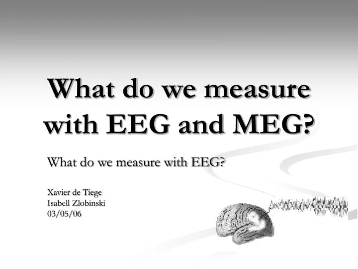
What do we measure with EEG and MEG?. What do we measure with EEG? Xavier de Tiege Isabell Zlobinski 03/05/06. Characteristics of the EEG. Localize neural electric activity using non-invasive measurements

E N D
What do we measure with EEG and MEG? What do we measure with EEG? Xavier de Tiege Isabell Zlobinski 03/05/06
Characteristics of the EEG • Localize neural electric activity using non-invasive measurements • Measures electric potential differences between pairs of scalp electrodes • Temporal precision • Rhythmic fluctuations in voltage • Standard time interval: cycles per second = hertz (Hz) • Amplitude in microvolts (µV)
Recording EEG • EPSP at apical dentritic trees of pyramid cells • Dentritic membrane depolarized • Potential difference cause a current flow through volume conductor from the nonexited membrane of the soma to the apical dentritic tree • Extracellular currents = secondary / volume currents
The EEG machine • 8 – 64 identical channels recording simultaneously from as many different pairs of electrodes • Electrodes & electrode board • Amplifiers • Filters • Pen & chartdrive (screen)
When do we use EEG? • Sleep research • Clinical diagnosis • Epileptic patients • Sleep disorders • Encephalopathies • Biophysiologic research (e.g. evoked potentials) • Cognitive research (e.g. ERPs) • …
Physiologic artifacts Eye movement Muscle activity ECG artifacts Skin artifacts Extraphysiologic artifacts Electrodes Alternating current (60 Hz) artifact Movements in the enviroment Artifacts
Event Related Potentials • Voltage fluctuations in cortex because of cognitive procedures or stimuli responses • Designed by summation & averaging of event related EEG parts • Waves described after polarity and latency (e.g. P300), method of release (mismatch negativity) and psychophysiological correlatives
ERPs – failures & limits • Artifacts possible • Difficult to analyse high complex cognitive procedures when stimuli need more time than 100 ms short ERP duration • between individuals very variable • depends on age • combined from several spacial and temporal overlapping components low specificity
Evoked Potentials • Record of low amplitude potentials evoked by different types of sensory stimulus • Voltage fluctuation is slow with a very small amplitude of the response (about 1/100 of spontaneous EEG activity) • high amplification is essential • Special computer averaging technic is required • clinical diagnostic, neurophysiologic & cognitive research • Visual evoked Potential • Brainstem auditory evoked Potential • Somatosensory evoked Potential
Sources • Duffy, Iyer, Surwillo (1989). Clinical Electroencephalography and Topographic Brain Mapping. Springer Verlag • S. Baillet, J.C. Mosher, R.M. Leahy. (2001). Electromagnetic Brain Mapping. IEEE Signal Processing Magazine. P 14-30. • www.dkgn.de • http://edoc.hu-berlin.de/dissertationen/eichholz-stephan-2004-10-22/HTML/chapter2.html • http://www.emedicine.com/neuro/topic678.htm