Download
1 / 24
240 likes | 384 Views
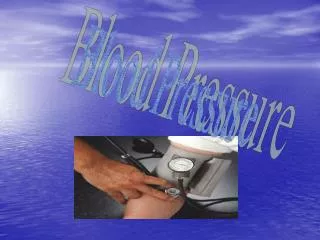
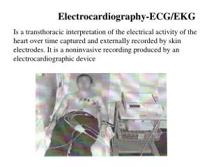
Bio102 Laboratory 6 Blood Pressure ECG (EKG). Objectives for Today’s Lab. Blood Pressure Become familiar with determining pulse rate Become familiar with taking a blood pressure reading and with using a sphygmomanometer (<- can you spell this? )

E N D
Bio102 Laboratory 6 Blood PressureECG (EKG)
Objectives for Today’s Lab • Blood Pressure • Become familiar with determining pulse rate • Become familiar with taking a blood pressure reading and with using a sphygmomanometer (<- can you spell this? ) • Correlate the effects of various experimental conditions on pulse and blood pressure • ECG • Acquire a better understanding of the theory behind the ECG • Record and analyze an ECG • Computers set up in lab • You will conduct a one-lead ECG on a lab partner • Identify the components (waves, intevals) of a normal ECG • Describe the phases of the cardiac cycle represented by each wave form on the ECG • Calculate heart rate from an ECG • Identify the beginning and end of the PR, QRS, and QT intervals
Location of Pressure Points Temporal artery Facial artery External Carotid artery Brachial artery Radial artery Pulse = # beats/30 sec X 2 Femoral artery Popliteal artery Posterior tibial artery Dorsalis pedis artery
Pulse in Lab Today • Use the radial artery of your partner, and use your index finger • Note the characteristics of the pulse • Regular, irregular • Strong, weak • Count number of pulses in 30 sec. and multiply by 2 to get the pulse rate • Do this sitting down, standing up, and after exercise
Heart Sounds • Lubb (S1) • first heart sound • occurs during ventricular contraction • A-V valves closing • Dupp (S2) • second heart sound • occurs during ventricular relaxation • semilunar valves closing ♥
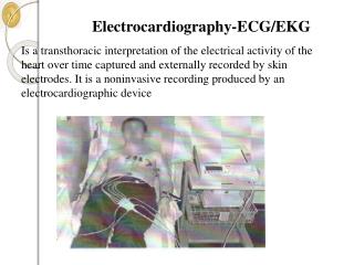
Heart Sounds Listening to the heart (auscultation) is done using a stethoscope (can you spell this?) Because sounds must pass through several layers of tissue, sounds are not heard directly above their point of origin S1 = “Lubb” (1 looks like L) S2 = “Dupp” (2 curved like D)
Heart Sounds Auscultate over the left sternal border about the level of the 3rd or 4th rib Find the point where you can hear the sounds the best, and use that Remember: lubb-dupp <pause> lubb-dupp <pause> …
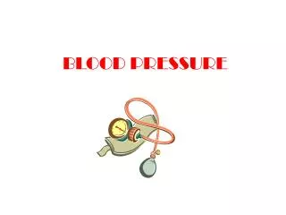
Auscultory Method of Determining Blood Pressure Use an average bp from two different readings from both arms. Do this standing up, sitting down, and after exercise Korotkoff sounds…
For the BP portion of today’s today… • If you need a bit of background and haven’t read it already, take a look at the first few pages of today’s handout • Also see Marieb’s Lab Manual, pp. 497-499
Great Book on Cardiovascular Physiology Excellent reference for cardiovascular physiology Author: R. E. Klabunde
A GREAT Book on ECGs This is you after reading the book…confident and no longer intimidated by ECGs!!
Electrocardiogram Millivolts Time PR Interval: 0.12 – 0.20 sec QT Interval: 0.20 – 0.40 sec QRS Interval: < 0.10 sec Figure from: Martini, Anatomy & Physiology, Prentice Hall, 2001
The Bipolar Limb Leads We will be using only Lead II today, so the right arm will be negative, and the foot will be considered positive
The Electrocardiogram A wave of the ECG will be positive (up) when the positive wave is moving toward the positive electrode
Electrocardiogram and Heart Events Right Arm(-) Left Foot (+) Recall that the left foot electrode is POSITIVE, so when the wave of depolarization is moving toward the left foot, we get a positive (up) deflection on the ECG
Electrocardiogram and Heart Events Right Arm (-) Remember that the T-wave represents repolarization of the ventricles. Left Foot(+)
ECG Animation… NYU ECG tutorial site… This is available on Links to Explore for Lab 6
The Paper Chart ECG Small boxes are usually 1 mm on each side Ten mm high represents 1 mV 0.04 sec 0.2 sec
The Paper ECG Chart How long is the PR interval on this strip? The QT interval? (See Marieb’s Lab Manual pg. 460 for explanation of these)
Calculation of Heart Rate • As shown in the sample ECG below, you will calculate your heart rate by measuring the time between 5-8 beats of your heart (4 beats shown below). • Then, to determine your heart rate from that, use the following proportion: • Beats measured (beats) * 60 seconds / Total time measured (sec) = Heart rate (bpm)
For the ECG portion of today’s lab… • If you haven’t read it already and need a bit of background, see Marieb’s Laboratory Manual pp. 457-460 • Run the ECGs for each member of your group by following the Procedure in your handout • Follow the instructions for the PowerLab ECG module in your handout • Be sure electrode placement is correct • Input the correct bioamp settings to reduce interference • Be sure to record values you measure on the EGG on the computer while you have them on the screen, and then transfer them to the ECG strip after you print it • Print each person’s ECG and directly on the printed ECG • Show calculations for the heart rate • Mark the PR, QRS, and QT intervals on hard copy with their times (that you recorded when they were on the screen) • Indicate the P, QRS, and T waves and what they mean
What you should do in lab today… • After EACH member of your group has an ECG recorded, be sure to SAVE the ECG into a file until the lab is over (just in case) • Select File | Save As (to prevent overwriting someone else’s file with your data) • Enter a file name using your first and last name with no spaces as the file name, e.g., SueCidal). • File is saved to the Desktop, so when you retrieve it later to analyze the data , LOOK ON THE DESKTOP!
Next Lab… • Lab 7 - Microcirculation • We will be examining the circulation in arterioles, capillaries, and venules • In a goldfish tail (real one; not the cracker) • Examining the effect of various substances/factors on circulation • See the handout online