Download
1 / 9
120 likes | 356 Views
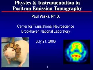
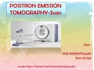
POSITRON EMISSION TOMOGRAPHY-Scan. Permadi Ardiansyah D411 05 093.

E N D
POSITRON EMISSION TOMOGRAPHY-Scan PermadiArdiansyah D411 05 093
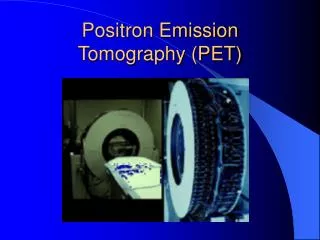
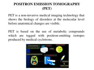
PET-scan adalahsuatualat yang digunakanuntukmendeteksipenyakitkanker. PET merupakansalahsatuhasildigarisdepanpengembanganradioisotopuntukduniakedokteran. PET menggunakanmetodevisualisasifungsitubuhmenggunakanradioisotoppemancar positron.
Keunggulan PET dari MRI dan CT-Scan Baik CT-Scan maupun MRI, kemampuanmendeteksikankerhanyaterbataspadaaspekanatomitubuhsaja. Sedangkanpada PET, aspekanatomidanmekanismekerja organ tubuh yang disebutmetobolismetubuhjugadapatdideteksialatini. Alatinibahkandapatmendeteksijeniskanker,tingkatkeganasan,lokasi,sertacararambatpenyakitkanker.
PRINSIP KERJA PET-CT Scan Perangkat PET secaragarisbesarterbagi 3: • Bagianproduksi flour-18 • Bagiansintesa 18FDG • Bagiankamera PET
Penggunaan PET diawalidenganmemproduksiradioisotop flour-18. Radiositopflour-18 diproduksidariisotop oksigen-18 menggunakansiklotron. Radioiaotop flour-18 yang telahdidapatkandigunakanuntukmensintesa 18FDG. Setelah 18FDG selesaidisiapkan, kemudiansegeradisuntikkankepasien. Jumlah yang disuntikkanantara 10 dan 20 milicurie.
Sebaranfluor-18 didalamtubuhdideteksidenganmemasukkantubuhkedalamrangkaiandetektorelektronikberbentukmelingkar. Dari hasilpendeteksianinidilakukan image reconstruction untukmendapatkangambaransebaran fluor-18 didalamtubuh. Perangkatkamera PET biasanyatelahdilengkapidengan program untukkeperluaninisehinggahasil image reconstruction dapatdiperolehdenganmudah.