Download
1 / 55
580 likes | 855 Views
Hormone Actions and Insulin Receptors By: Netee Papneja, PGY5. Netee Papneja PGY 5, Endocrinology. Objectives:. Introduction to hormones: Definition Classification Structure Synthesis and release Function Hormone receptors: Classification Brief overview of each class

E N D
Hormone Actions and Insulin ReceptorsBy: Netee Papneja, PGY5 Netee Papneja PGY 5, Endocrinology
Objectives: • Introduction to hormones: • Definition • Classification • Structure • Synthesis and release • Function • Hormone receptors: • Classification • Brief overview of each class • Insulin and insulin receptors: • General information • Mechanism of action
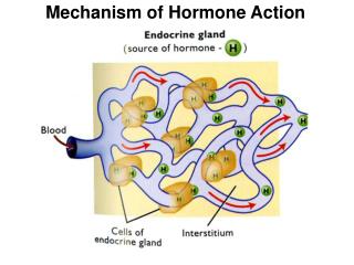
What is a hormone? • A hormone (from Greek ''ὁρμή'' - "impetus") is a chemical released by one or more cells that affects cells in other parts of the organism. • a chemical messenger that transports a signal from one cell to another • Only a small amount of hormone is required to alter cell metabolism
Endocrine hormones - secreted (released) directly into the bloodstream • Exocrine hormones - secreted directly into a duct, and from the duct they either flow into the bloodstream or they flow from cell to cell by diffusion in a process known as paracrine signalling.
The physiologic functions of hormones can be divided into three general areas: • Growth and differentiation: • Multiple hormones and nutritional factors • Reproduction: • The stages of reproduction include: • Sex determination during fetal development • Sexual maturation during puberty • Conception, pregnancy, lactation, and child-rearing • Cessation of reproductive capability at menopause
Maintenance of homeostasis • T4 controls about 25% of basal metabolism in most tissues • Cortisol exerts a permissive action for many hormones in addition to its own direct effects • PTH regulates calcium and phosphorus levels • Vasopressin regulates serum osmolality by controlling renal free water clearance • Mineralocorticoids control vascular volume and serum electrolyte (Na+, K+) concentrations • Insulin maintains euglycemia in the fed and fasted states
Communication system • Optimal coordination and communication between organ systems is required to sustain homeostasis • A complete communication system needs: • A cell that produces the signaling molecule (the hormone, which is sometimes called the ligand) • A target cell with a specific receptor that can bind the signal with high affinity and produce a desired effect.
Receptors: • molecules that hormones bind to in order to exert their effects • Characteristics of receptors: • Proteins or glycoproteins • Able to distinguish their hormone from other molecules that may have very similar structures
Characteristics of receptors: • Bind to the hormone, or ligand, even at exceedingly low concentrations • Undergo a conformational change when bound to the hormone • Catalyze biochemical events or transmit changes in molecular conformation to adjacent molecules that produce a biochemical change
General Classification: • Membrane receptors • Nuclear receptors
Nuclear Receptors • Change the degree of gene expression • Can be located in the cytoplasm or in the nucleus • Steroid receptors • In the cytoplasm • steroid diffuses through the membrane and binds to its receptor • It then dissociates from proteins and translocates to the nucleus, where another steroid-receptor complex binds to it to form a dimer of steroid-receptor complexes, which exposes the DNA-binding site and, at this point, becomes active. • Thyroid hormone receptors • Are already in the nucleus and bound to the target genes • There are inactivating thyroid binding proteins that dissociate once the hormone has bound, allowing the hormone-receptor complexes to cause changes in gene expression
Membrane Receptors • Activated through the binding of peptide hormones and catecholamines • The ligand (hormone), or first messenger, binds to its receptor and causes activation of a second messenger system, which is mediated with intracellular signalling molecules (through phosphorylation reactions)
Membrane receptors can be classified according to the molecular mechanisms by which they accomplish their signaling function: • Ligand-gated ion channels (e.g., nicotinic acetylcholine receptor) • Receptor tyrosine kinases (e.g., receptors for insulin and insulin-like growth factor I [IGF-I]) • Receptor serine/threonine kinases (e.g., receptors for activins and inhibins) • G protein–coupled receptors (e.g., receptors for adrenergic agents, muscarinic cholinergic agents, glycoprotein hormones, glucagon, and parathyroid hormone) • Cytokine receptors (e.g., receptors for growth hormone, prolactin, and leptin)
Ligand-gated ion channels • Receptor tyrosine kinases • Receptor serine/threonine kinases • Receptor guanylate cyclase • Bifunctional molecules that can bind hormone as well as serve as effectors by functioning either as ion channels or as enzymes.
G protein–coupled receptors • Cytokine receptors • Have the ability to bind the hormone but must recruit a separate molecule to catalyze the effector function.
G protein-coupled receptors • Span the membrane seven times, with the receptor extracellularly and regions that activate a G protein intracellularly • Three components: α, β and γ subunits • Ligand binding to the receptor results in a conformational change that causes the α subunit to exchange a GDP molecule for that of a GTP molecule • This causes the GTP-bound α subunit to dissociate from the βγ complex and act at an effector (usually an enzyme, but sometimes an ion channel or other protein). • The α subunit hydrolyses GTP back to GDP to terminate the process.
Receptor Tyrosine Kinases: • Have several structural features in common: • an extracellular domain containing the ligand-binding site • a single transmembrane domain • an intracellular portion that includes the tyrosine kinase catalytic domain
100 receptor tyrosine kinases sequenced in the human genome • Classified into 16 subfamilies based on the extracellular domain • Mediate the actions of many ligands: • insulin, • epidermal growth factor (EGF), • platelet-derived growth factor (PDGF) • vascular endothelial cell–derived growth factor.
Receptor activation: Role of Dimerization: • Plays a central role in the activation of most receptor tyrosine kinases. • The molecular mechanisms differ from receptor to receptor
Examples of mechanisms of dimerization: • Ligand has two subunits, each binds to a receptor (dimeric ligand) • Two receptor-binding sites on a single molecule of ligand • Pre-existing receptor dimers, undergo conformational change and become activated when bound to ligand
After dimerization, receptor tyrosine kinases undergo conformational change in the kinase domain • Mainly due to autophosphorylation of the “activation loop” • Ultimately leads to activation of the receptor to phosphorylate other proteins
Termination of signal: • Receptor-Mediated Endocytosis: • Protein Tyrosine Phosphatases: • Serine/Threonine Kinases
Receptors that Signal through Associated Tyrosine Kinases • Members of the cytokine family of receptors resemble receptor tyrosine kinases in their mechanism of action • Instead of the tyrosine kinase being intrinsic to the receptor, enzymatic activity resides in a protein that associates with the cytokine receptor. • Ligand binding to the cytokine receptor activates the associated kinase.
Insulin Physiology • Insulin – key regulator of glucose, protein, fat homeostasis • 51-amio acid anabolic hormone made of 2 peptide chains connected by 2 disulfide chains
Insulin Biosynthesis • Pre-proinsulin(PPI) synthesized from the • Insulin gene • PPI into the endoplasmic reticulum • by the signal sequence, where it folds into • a proper conformation that is stabilized • by three disulfide bonds (SS) • The signal sequence is removed • and proinsulin is further processed in the • Golgi apparatus, where the C-peptide is • removed and packaged with insulin • for secretion
Proinsulin • A single chain of 86 amino acids • Small amount escapes from pancreas uncleaved and circulates in serum • Metabolized in kidney instead of liver therefore 3 to 4-times the half life of insulin • 12-20% of circulating “insulin” that we measure in the fasting state • Has 7-8% of insulin’s biologic activity
C-Peptide • 31 amino acid peptide • No known biologic activity • Excreted by kidney • Half-life 3-4-times that of insulin
Insulin • Processed and secreted by pancreatic B cells of the islets of Langerhans • Transported via portal circulation to hepatic vein to the liver • 50% of insulin is removed by a single pass through the liver, remainder of it by kidneys • Circulatory half-life of 3-5 minutes
Insulin Secretion • Pancreas secretes about 30 units per day • Release of insulin occurs at a basal rate and in short lived large bursts with glucose load • Basal insulin secretion occurs during fasting/resting states to inhibit hepatic glycogenolysis, ketogenesis, and gluconeogenesis • 40% of total insulin output/24h
Insulin Secretion • Bolus insulin occurs when plasma glucose levels are >4.4-5.6mmol/L after meals • Bolus insulin released in 2 phases • First phase: initial transient surge • Second phase: prolonged steady increase • Increased levels start 8-10 minutes after eating and peaks in 30-45 minutes
INSULIN RELEASE • Glucose into pancreatic cell via GLUT-2 transporters • Glucose metabolism generates ATP • Glucokinase = key enzyme, glucose concentration dependent, catalyzes the conversion of Glucose to G6P • ATP displaces ADP from open ATP sensitive K channels • Bound ATP causes causes channels to close, restricting efflux of K = deploarizing the cell • Depolarlization opens Voltage gated calcium channels triggering exocytosis of insulin vesicles
Insulin receptor • Most body cells (hepatocytes, fat, muscle cells) have insulin receptors • Composed of: • Two alpha subunits and two beta subunits linked by disulfide bonds • It is a transmembrane receptor that is activated by insulin, IGF-1, IGF-II, and belongs to class of tyrosin kinase receptors • a kinase is a type of enzyme that transfers phosphate groups from high-energy donor molecules, such as ATP to specific target molecules phosphorylation. • Kinase enzymes that specifically phosphorylate tyrosine amino acids are termed tyrosine kinases.
Mitogenic functions: mediated via the mitogen-activated protein kinase (MAP kinase) pathway. • Metabolic actions: mediatedby phosphatidylinositol-3-kinase (PI-3K) pathway
PI-3K-signaling pathway is responsible for: • Translocation of GLUT-4 containing vesicles to the surface • Increasing GLUT-4 density on the membrane and rate of glucose influx • Promoting glycogen synthesis via activation of glycogen synthase • Promoting protein synthesis and lipogenesis, while inhibiting lipolysis
Insulin Receptors • Insulin binds to α subunits of insulin receptor tyrosine kinase and causes shape changes communicated to the intracellular β subunits and cause it to bind ATP and autophosphorylate • This then allows other intracellular proteins to bind to the intracellular domain of the receptor, and become phosphorylated and generate their actions. • A cascade of phosphorylations and shape/activity changes START
Signal transduction pathways: • IRS (insulin receptor substrates) binding sites for PI3K (phosphatidylinositol-3-kinase) activates Akt/PKB (Protein Kinase B) and the aPKC (Protein Kinase C) cascades
Activated Akt/PKB induces glycogen synthesis through inhibition of GSK-3 (Glycogen synthase kinase 3); protein synthesis via mTOR (mammalian target of rapamycin) and gluconeogenesis via FOXO-1 (Forkhead box protein O1)
Glucose uptake… • Insulin stimulates glucose uptake via translocation of GLUT4 vesicles to the plasma membrane • Activation of PKB and PKC-λ lead to translocation of GLUT4 molecules to the cell surface resulting in increased glucose uptake
Insulin signaling also has growth and mitogenic effects, which are mostly mediated by the Akt cascade as well as by activation of the Ras/ MAPK pathway