Download
1 / 95
1.06k likes | 1.45k Views
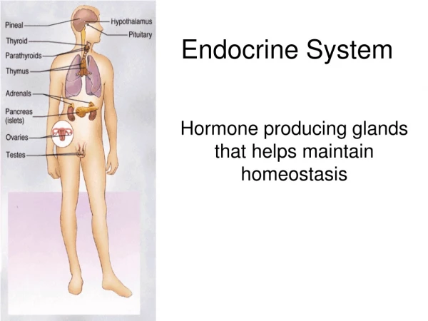
Endocrine system. Hormone. The term hormone is derived from a Greek verb meaning – to excite or arouse Hormone is a chemical messenger that is released in one tissue (endocrine tissue/gland) and transported in the bloodstream to reach specific cells in other tissues

E N D
Hormone • The term hormone is derived from a Greek verb meaning – to excite or arouse • Hormone is a chemical messenger that is released in one tissue (endocrine tissue/gland) and transported in the bloodstream to reach specific cells in other tissues • Regulate the metabolic function of other cells • Have lag times ranging from seconds to hours • Tend to have prolonged effects • Hormone actions must be terminated – how?
Endocrine versus Nervous system • Both use chemical communication • Both are being regulated primarily by negative feedback • Released in synapse • Close to target cells • Signal to release by action potential • Short live effect • Crisis management Neurotransmitters Hormones • Released to bloodstream • Can be distant from target cells • Different types of signal • Long term effect • Ongoing processes
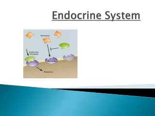
Intercellular communication types • Autocrine - the cell signals itself through a chemical that it synthesizes and then responds to. Autocrine signaling can occur: • solely within the cytoplasm of the cell or • by a secreted chemical interacting with receptors on the surface of the same cell • Paracrine - chemical signals that diffuse into the area and interact with receptors on nearby cells (cells within the same tissue). • Endocrine - the chemicals are secreted into the blood and carried by blood and tissue fluids to the cells they act upon. http://users.rcn.com/jkimball.ma.ultranet/BiologyPages/H/Hormones.html
Control of Hormone Release • Blood levels of hormones: • Are controlled by negative feedback systems • Vary only within a narrow desirable range • Hormones are synthesized and released in response to: • Humoral stimuli • Neural stimuli • Hormonal stimuli
Humoral Stimuli • Secretion of hormones in direct response to changing blood levels of ions and nutrients • Example: concentration of calcium ions in the blood • Declining blood Ca2+ concentration stimulates the parathyroid glands to secrete PTH (parathyroid hormone) • PTH causes Ca2+ concentrations to rise and the stimulus is removed
Neural Stimuli • Neural stimuli – nerve fibers stimulate hormone release • Preganglionic sympathetic nervous system (SNS) fibers stimulate the adrenal medulla to secrete catecholamines Figure 16.5b
Hormonal Stimuli • Hormonal stimuli – release of hormones in response to hormones produced by other endocrine organs • The hypothalamic hormones stimulate the anterior pituitary • In turn, pituitary hormones stimulate targets to secrete still more hormones
Classes of Hormones – by chemical structure • Hormones can be divided into three groups • Amino acid derivatives • Peptide hormones • Lipid derivatives
Amino Acid Derivatives Derivatives of Tyrosine: Thyroid hormones Catecholamines Epinephrine, norepinephrine Derivatives of Tryptophan: Dopamine, serotonin, melatonin
Peptide Hormones 3 groups – glycoproteins, short peptides and small proteins Glycoproteins Proteins are more than 200 amino acids long and have carbohydrate side chains Thyroid-stimulating hormone (TSH) Luteinizing hormone (LH) Follicle-stimulating hormone (FSH)
Peptide Hormones Short chain polypeptides Antidiuretic hormone (ADH) and oxytocin (OXT) (each 9 amino acids long) Small proteins Growth hormone (GH; 191 amino acids) and prolactin (PRL; 198 aminoacids) from the pituitary gland Includes all hormones secreted by: Hypothalamus, heart, thymus, digestive tract, pancreas, and posterior lobe of the pituitary gland, as well as several hormones produced in other organs
Lipid Derivatives – include Eicosanoids and steroids Eicosanoids- derived from arachidonic acid, a 20-carbon fatty acid Paracrine factors prostaglandins - involved primarily in coordinating local cellular activities Steroid hormones - derived from cholesterol Released by: The reproductive organs (androgens by the testes in males, estrogens and progestins by the ovaries in females) The cortex of the adrenal glands (corticosteroids) The kidneys (calcitriol)
Distribution of Hormones in bloodstream • Hormones that are released into the blood are being transported in one of 2 ways: • Freely circulating • Bound to transport protein
Distribution of Hormones in bloodstream • Freely circulating (most hormones) • Hormones that are freely circulating remain functional for less than one hour and some as little as 2 minutes • Freely circulating hormones are inactivated when: * bind to receptors on target cells * being broken down by cells of the liver or kidneys * being broken down by enzymes in the plasma or interstitial fluid • Bound to transport proteins – thyroid and steroid hormones (>1% circulate freely) • Remain in circulation longer
Hormones: Classification Table 7-1
Target Cell Specificity • Hormones circulate to all tissues but only activate cells referred to as target cells • Target cells must have specific receptors to which the hormone binds • These receptors may be intracellular or located on the plasma membrane
Interaction of Hormones at Target Cells • Three types of hormone interaction • Permissiveness – one hormone cannot exert its effects without another hormone being present • For example, thyroid hormone increases the number of receptors available for epinephrine at the latter's target cell, thereby increasing epinephrine's effect at that cell. Without the thyroid hormone, epinephrine would only have a weak effect • Synergism – more than one hormone produces the same effects on a target cell • Antagonism – one or more hormones opposes the action of another hormone
Target Cell Activation • Hormone exert their effects on target cells at very low bloodconcentrations (ng-10-9 gr; pg-10-12 gr) • Target cell activation depends on three factors • Blood levels of the hormone • Relative number of receptors on the target cell • The affinity of those receptors for the hormone • The time required to effect target cells depends on the hormone - some influence immediately and some (steroids; why?) require hours or days • Hormone effect duration also varies and can range between seconds to hours
Receptors number on target cell • down regulation – the presence of the hormone induces a decrease in the receptors concentration; • high levels of hormone – cell less sensitive • Up regulation – absence of the hormone induces the increase in receptors concentration; • Low levels of hormone – cell more sensitive • In most systems the maximum biological response is achieved at concentrations of hormone lower than required to occupy all of the receptors on the cell (spare receptors). • Examples: • insulin stimulates maximum glucose oxidation in adipocytes with only 2-3% of receptors bound • LH stimulates maximum testosterone production in Leydig cells when only 1% of receptors are bound
Receptors for hormones are located: • In the cell membranes of target cells • In the cytoplasm or nucleus
Mechanisms of Hormone Action • Two mechanisms, depending on their chemical nature • Water-soluble hormones (all amino acid–based hormones except thyroid hormone) • Cannot enter the target cells • Act on plasma membrane receptors • Coupled by G proteins to intracellular second messengers that mediate the target cell’s response • Lipid-soluble hormones (steroid and thyroid hormones) • Act on intracellular receptors that directly activate genes
Receptors on the cell membrane • Hormones do not induces changes in cell activity directly but via the induction of the appearance and action of other agents • Hormones are referred to as first messengers and the agents that are activated by the hormones are called second messengers. • All amino-acid hormones (with exception of the thyroid hormone) exert their signals through a second messenger system: • cAMP • PIP
Links the first messenger (hormone) and the second messenger Hormone Protein receptor G protein (inactive) G protein activated The actions of second messengers for hormones that bind to receptors in the plasma membrane Effects on Ca2+ Levels Effects on cAMP Levels Some G proteins use Ca2+ as a second messenger. Many G proteins, once activated, exert their effects by changing the concentration of cyclic-AMP, which acts as the second messenger within the cell. Ca2+ Hormone Hormone Hormone Protein receptor Protein receptor Protein receptor Enhanced breakdown of cAMP Increased production of cAMP G protein activated G protein activated G protein activated Opening of Ca2+ channels Release of stored Ca2+ from ER or SER Acts as second messenger cAMP cAMP ATP AMP Ca2+ Ca2+ Ca2+ acts as second messenger Ca2+ Reduced enzyme activity Calmodulin Opens ion channels Activates enzymes Activates enzymes If levels of cAMP increase, enzymes may be activated or ion channels may be opened, accelerating the metabolic activity of the cell. In some instances, G protein activation results in decreased levels of cAMP in the cytoplasm. This decrease has an inhibitory effect on the cell. The calcium ions themselves serve as messengers, generally in combination with an intracellular protein called calmodulin. Figure 16.2 1
Receptors on the cell membrane • Second messengers function as enzyme activator, inhibitor or cofactor • A small number of hormone molecules induce the appearance and activity of many 2nd messenger molecules – amplification • one single hormone can induce the activation of more than one 2nd messenger • Activation of a 2nd messenger can start a chain of reactions – receptor cascade
Amino Acid-Based Hormone Action: cAMP Second Messenger • Hormone (first messenger) binds to its receptor, which then binds to a G protein • The G protein is then activated • Activated G protein activates the effector enzyme adenylate cyclase • Adenylate cyclase generates cAMP (second messenger) from ATP • cAMP activates protein kinases, which then cause cellular effects
Extracellular fluid 1 Hormone (1st messenger)binds receptor. Adenylate cyclase G protein (GS) 5 cAMP acti-vates proteinkinases. Receptor Activeproteinkinase GDP Inactiveprotein kinase 2 3 4 Receptoractivates Gprotein (GS). G proteinactivatesadenylatecyclase. Adenylatecyclaseconverts ATPto cAMP (2ndmessenger). Hormones thatact via cAMPmechanisms: Triggers responses oftarget cell (activatesenzymes, stimulatescellular secretion,opens ion channel,etc.) GlucagonPTHTSHCalcitonin EpinephrineACTHFSHLH Cytoplasm Figure 16.2, step 5
Amino Acid-Based Hormone Action: PIP-Calcium • Hormone binds to the receptor and activates G protein • G protein binds and activates phospholipase • Phospholipase splits the phospholipid PIP2 into diacylglycerol (DAG) and IP3 (both act as second messengers) • DAG activates protein kinases; IP3 triggers release of Ca2+ stores • Ca2+ (third messenger) alters cellular responses
Amino Acid-Based Hormone Action: PIP Mechanism Extracellular fluid Hormone DAG 1 4 5 Active protein kinase C 2 3 PIP2 GTP GTP Receptor Gq Inactive protein kinase C GDP GTP IP3 Phospholipase C Catecholamines TRH ADH GnRH Oxytocin Triggers responses of target cell 5 Endoplasmic reticulum 6 Cytoplasm Ca2+ Ca2+- calmodulin Figure 16.3
Steroid Hormones: Action 1 Most hydrophobic steroids are bound to plasma protein carriers. Only unbound hormones can diffuse into the target cell. Blood vessel Steroid hormone Cell surface receptor 2a Rapid responses 1 2 Steroid hormone receptors are in the cytoplasm or nucleus. 2 Protein carrier Nucleus 2a Some steroid hormones also bind to membrane receptors that use second messenger systems to create rapid cellular responses. Cytoplasmic receptor Nuclear receptor DNA Interstitial fluid 3 The receptor-hormone complex binds to DNA and activates or represses one or more genes. 3 Endoplasmic reticulum Transcription produces mRNA Cell membrane 4 Activated genes create new mRNA that moves back to the cytoplasm. 5 4 New proteins Translation 5 Translation produces new proteins for cell processes. Figure 7-7, steps 1–5
http://arbl.cvmbs.colostate.edu/hbooks/pathphys/endocrine/moaction/change.htmlhttp://arbl.cvmbs.colostate.edu/hbooks/pathphys/endocrine/moaction/change.html
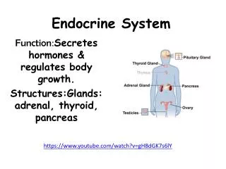
How will we approach the endocrine system? • We will group them according to their function in the body: • Hormones that control blood glucose levels • Hormones that control minerals and water balance • Hormones that are involved in growth and metabolism • Hormones and the reproductive system
Pancreas structure Exocrine pancreas (99% of volume) Cells (pancreatic acini) forming glands and ducts that secrete pancreatic fluid and enzymes with digestive function Endocrine pancreas (1%) Small groups of cells scattered in clusters (pancreatic islets) that secrete hormones
Pancreas – islets of Langerhans cells • The islets contain two major cell types: • Alpha () cells that produce glucagon • Beta () cells that produce insulin • The islets also contain • Delta cells – produce a peptide hormone identical to GHinhibiting hormone (GH-IH). That hormone suppresses the release of glucagon and insulin and slows food absorption and digestive enzyme secretion • F cells – Produce the hormone pancreatic polypeptide (pp) that inhibits gallbladder contractions and regulate the production of some pancreatic enzymes
How does the body control blood glucose levels • Blood Glucose Levels are controlled mainly by insulin and glucagon • When levels rise • Beta cells secrete insulin, stimulating transport of glucose across plasma membranes • When levels decline • Alpha cells release glucagon, stimulating glucose release by liver
Insulin • A 51-amino-acid protein consisting of two amino acid chains linked by disulfide bonds • Insulin is released when glucose levels exceed normal levels (70-110 mg/dl) http://www.chemistryexplained.com/images/chfa_02_img0437.jpg
Effects of Insulin Binding to its receptors • Insulin facilitates entry of glucose cells by binding to a membrane receptor • The complex insulin-receptor make a specific carrier protein (GLUT4) available • Once at the cell surface, GLUT4 facilitates the passive diffusion of circulating glucose down its concentration gradient into cells. • Receptors for insulin are present in most cell membranes (insulin-dependant cells) • Cells that lack insulin receptors are cells in the brain, kidneys, lining of the digestive tract and RBC (insulin-independent cells). • Those cells can absorb and utilize glucose without insulin stimulation.
Effects of Insulin • Acceleration of glucose uptake as a result from an increase of the number of glucose carrier proteins • Acceleration of glucose utilization and increased ATP production • Stimulation of glycogen formation in the liver and muscle cells • Inhibits glycogenolysis (break down of glycogen) and gluconeogenesis (glucose building) • Stimulation of amino acid absorption and protein synthesis • Stimulation of triglyceride formation in adipose tissue • As a result glucose concentration in the blood decreases
Glucagon • Released by alpha cells • A 29-amino-acid polypeptide hormone that is a potent hyperglycemicagent • it promotes: • Glycogenolysis– the breakdown of glycogen to glucose in the liver and skeletal muscle • Gluconeogenesis – synthesis of glucose from lactic acid and noncarbohydrates in the liver • Release of glucose to the blood from liver cells • breakdown of triglycerides in adipose tissue
Adrenal (Suprarenal) Glands • Structurally and functionally, they are two glands in one • Adrenal medulla – neural tissue; part of the sympathetic nervous system • Adrenal cortex - three layers of glandular tissue that synthesize and secrete corticosteroids
Adrenal Cortex • Synthesizes and releases steroid hormones called corticosteroids • Different corticosteroids are produced in each of the three layers • Zonaglomerulosa – glomerulus- little ball. Secretes mineralocorticoids – main one aldosterone • Zonafasciculata– glucocorticoids (chiefly cortisol) • Zonareticularis – gonadocorticoids (chiefly androgens)
Zonafasciculata - Glucocorticoids (Cortisol/hydrocortisone) • Main hormones secreted are the Cortisol/hydrocortisone and small amounts of corticosterone • Glucocorticoids often called the body’s stress hormones • While adrenaline is responsible for rapid metabolic responses the glucocorticoids are responsible for long-term stress: • Glucocorticoids accelerate the rates of glucose synthesis and glycogen formation – especially in the liver • Adipose tissue responds by releasing fatty acids into the blood and the tissues start to utilize fatty acids as source of energy - glucose-sparing effect (GH has similar effect and will be discussed later) • Clucocorticoids also have anti-inflammatory effect – inhibit the activities of WBC (use?)
Diabetes Mellitus (DM) • Two types: • Type I results from the destruction of beta cells and the complete loss of insulin (hypoinsulinemia) • Type II is the most common type (90%) and is a result of decrease sensitivity of cells to insulin (insulin resistance). Type II is accompanied by hyperinsulinemia (what is that? Why?). • Type II is associated with excess weight gain and obesity but the mechanisms are unclear. • Other reasons that were associated with type II diabetes: pregnancy, polycystic ovary disease, mutations in insulin receptors and others
Type 1 and Type 2 Diabetes Mellitus Table 24.1
Diabetes Mellitus (DM) effects • Increase in blood glucose due to diabetes causes • Increase in glucose loss in urine • Dehydration of cells – since glucose does not diffuse through cell membrane and there is an increase in osmotic pressure in the extracellualr fluid. • In addition, the loss of glucose in the urine causes osmotic diuresis - decrease in water reabsorption in the kidney. • The result is • Polyuria – huge urine output and dehydration. • Polydipsia – excessive thirst
Diabetes Mellitus (DM) effects • Polyphagia – excessive hunger and food consumption because cells are starving • Damage to blood vessels and poor blood supply to different tissues • Increase use of lipids as a source of energy by the cells and increase release of keto bodies – ketosis and changes of blood pH (acidosis). That leads to increased respiratory rate