Download

1 / 33
370 likes | 619 Views
Physiological changes in pregnancy. Dr. Wulan Margasari Soemardji, SpOG. The major maternal physiological adaptation to pregnancy. 1-Systemic changes: -volume homeostasis. -blood -cardio vascular system. 2-Respiratory changes. 3-urinary tract and renal function. 4-Alimentary tract.

E N D
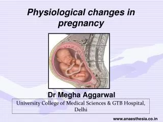
Physiological changes in pregnancy Dr.Wulan Margasari Soemardji, SpOG
The major maternal physiological adaptation to pregnancy 1-Systemic changes: -volume homeostasis. -blood -cardio vascular system. 2-Respiratory changes. 3-urinary tract and renal function. 4-Alimentary tract. 5-Reproductive organs. 6-endocrinological changes.
systemic changes • volume homeostasis: • fluid retention is the most fundamental systemic changes of normal pregnancy. • the total blood volume is increased during pregnancy 30%. • the most marked expansion occurs in extra cellular volume (ECV) with some increase in intra cellular water.
The factors contributing including: • Increase sodium retention. • Decrease in plasma osmotic pressure. • Decrease in thirst threshold. • Resetting of osmostate. • Decrease in plasma oncotic pressure.
Blood: The marked increase in plasma volume associated with normal pregnancy causes dilution of many circulating factors. Hematological changes Decrease in: • red cell count. • hemoglobin concentration. • haematocrit. • plasma folate concentration. Increase in : • white cell count. • erythrocyte segmentation rate . • fibrogen concentration.
Cardio vascular changes: Earliest changes is periphral vasodilatation Results in decreased systemic vascular resistence→ ↑CO 6 L/ min. Max. (22-28)wks. • heart rate increase (10-20%). • stroke volume increase (10%). • cardiac out put increase (30-50%). • Mean arterial blood pressure decrease (10%).- • Peripheral resistance decrease (35%).-
normal changes in heart sounds during pregnancy: • increase loudness of both S1 & S2. • >95% develop systolic murmur which disappears after delivery. • 20% have a transient diastolic murmur. • 10% develop continues murmur due to increase mammary blood flow. • ectopics • Relative tachy cardia • collapsing pulse
Respiratory changes • increaseO2 demand by 20 %. • ↑tidal volume with normal respiratory rate. • ↑po2 and ↓pco2 withcompensatory ↓HCO3(mild compensated respiratory alkalosis).
ventilatory changes: • thoracic anatomy changes. • tidal volume increases. • vital capacity increase. • functional residual capacity decrease.
The urinary tract and renal function • blood flow increase (60-70%). • glomerular filtration increased (50%). • clearance of most substances is enhanced. • plasma creatinine ,urea,urate are reduced. • glycoseuria is normal.
Alimentary system changes • the gums becomes spongy. • the lower oesophageal sphincter is relaxed (hurt burn). • gastric secretion is reduced. • the intestinal musculature is relaxed (constipation).
in early pregnancy uterine growth result from both hyperplasia and hypertrophy while later hypertrophy accounts for most of increase. • it weight one kilo gram at term( in pre pregnancy 50-60 grams • as the pregnancy advanced the uterus divided into upper and lower uterine segment the lower uterine segment composed of lower part of uterus and the upper cervix composed mainly from connective tissue because of this the lower uterine segment becomes stretched in late pregnancy.
the cervix: • the cervix becomes softer and swollen in pregnancy with the result columnar epithelium lining cervical canal becomes exposed to vaginal secretion. • oestradiol stimulate growth of columnar epithelial of the cervical canal so it becomes violte and is called ectropine. • the mucus gland becomes distended and secrete mucus which forms a mucus plug that is expelled in labour as the show. • prostaglandins and collagenase especially in last weeks of pregnancy act on collagen fiber make cervix more softer.
the vagina : • the vaginal mucosa becomes thicker during pregnancy. • the vaginal discharge during pregnancy increased due to increase desquamation of the superficial vaginal mucosal cells
D-breasts and lactation : • the earliest changes is a swelling of the breast tissue. • oestrogen leads to increase in number of glandular ducts. • progesterone leads to proliferation of glandular epithelium of the alveoli. • prolactine leads to active secretion of milk after birth.
Endocrinological changes: • prolactine concentration increases markedly but act after delivery. • human growth hormone is suppressed . • insulin resistance develop. • thyroid function changes little. • trans placental calcium transport is enhanced. • corticosteroid concentration increased. • aldesterone concentration increased. • angiotensin and renine increased
Hormones produced within uterus human chorionic gonadotrophin (HCG): • it is secreted by trophoblast and can be detected in serum 10 days after conception (RIA). • there is high level of circulating HCG in early pregnancy (to provide a suitable environment for implantation and development). • to support corpus luteum secretion of oestrogen and progesterone in the first trimester until the placenta becomes able to produce these hormone. • the peak level normally occur in the 12th week .
constant level of HCG in late pregnancy is useful in: • controlling placental secretion of Estrogen progesterone. • suppressing maternal immune system against fetus. • the human chorionic gonadotrophine normally disappear from urine 7-10 days after delivery of placenta.
human placental lactogen • it is secreted by syncytotrophoblast. • It is level increase when the level of HCG start to drop . • HPL has no effect on fetus. • HPL effect on : 1-the breast: • mammary growth during pregnancy. • produce of colostrums. • milk production lactation.
2-protiens: • HPL stimulate protein synthesis at cellular level. 3-carbohydrate: • stimulate insuline secretion . • inhibit insulin action. 4-fat: HPL mobilize fat from body store (lypolysis) lead to increase maternal blood glucose and maternal tissue can not utilze the glucose so the glucose will be available for fetus.
Estrogen • it is produce by corpus luteum in early pregnancy. • it is produce by placenta in late pregnancy. • fetus (liver and adrenal ) provide certain enzyme which are lack in placenta. role of estrogen: • On connective tissue: estrogen leads to polymerization of mucopoly saccarides of the ground substance leads to loose connective tissue mainly in the cervix. • On the protein: estrogen stimulate directly RNA synthesis lead to protein synthesis.
progesterone • it is production same as estrogen. • it has effect on smooth muscle leads to decrease muscle excitability leads to muscle relaxation mainly in uterus.
Thyroid function • increase thyroid binding globulin. • increase bound form of T3,T4. • no change in free form of T3,T4. So no evidence to support what previously thought to be physiological such as increase in size of thyroid gland , increase BMR, body temperature, heart rate.
Diagnosis of pregnancy • History: symptoms. • Examination: signs. • Investigation : pregnancy test and ultrasound.
symptoms of pregnancy 1-Amenorrhoea: abrupt cessation of menses in a woman with regular cycle is highly suggestive. 2-breast symptoms: tenderness and fullness may be noticed . 3-frequency of micturation : pressure on the urinary bladder by enlarging uterus.
4-nausea with or without (morning sickness). 5-abdominal enlargement. 6-fetal movement: • quickening is the first feels fetal movement PG at (18-20wks). • Multi para at (16-18wks).
signs of pregnancy 1-breasts signs: • enlargement and increase pigmentation of the nipple. • increased pigmentation in the areola (areola). • formation of secondary areola. • montgomery areola or tubercle: • small tubercles 12-20 at the periphery of primary areola appear at 8th week due to active sebaceous gland. • prominent vein on the surface. • colostrum at 16th week is reliable in primigravida.
2-skin signs: • linear nigra. • stria gravidarum. • chloasma.
3-genital tract signs: • bluish discolouration of the vulva. • genital tract becomes more soft and warm. • Uterine changes: • uterus becomes abdominal organs at 12th week. • uterus becomes rounded (globular) instead of flatten in antero posterioly. • uterus becomes soft due to increase vascularity.
4-signs due to presence of the fetus: • fetal heart sounds: • after 12 weeks fetal heart heard with fetal sonicaid. • after 24th week fetal heart heard with fetal stethoscope. • FHR 120-160 beats/minuts. • funic soufflé:heard when fetal steatoscope lie directly over umbilical cord it is soft blowing murmur synchronous with fetal heart sounds. • palpitation of fetal parts from 24th weeks. • fetal movement:may felt during palpation. • Braxton hicks sign:irregular painless contraction palpable at 20th week.
investigation 1-pregnancy tests: • a pregnancy tests detects human chorionic gonadotrophine(HCG) in mother urine or serum. urine tests: agglutation inhibition (day 35 after LNMP). • standard HCG is adsorbed on particles or cells in suspension.. • anti serum (Ab) and some of patient urine is added. • if urine contains HCG it will combine with the antibody and thus prevents it from binding and agglutinating the particles.
if urine containing no HCG anti body binds adjacent particles thus causing agglutination. • the test can be carried out on slides or in tubes. blood tests (day 10 after implantation): • radio immune assay (RIA). • Enzyme-linked immuno assay (Elisa): • Can detect levels as low as 0.1-0.3 iu/l • Can detect pregnancy before the patient missed period.
Ultrasonography • 4 weeks: pregnancy sac with decidual reaction . • 5 weeks: yolk sac. • 6 weeks: fetal echo. • 6-7 weeks : presence of fetal heart. • 9 weeks :fetal morphology.