Download
1 / 49
530 likes | 732 Views
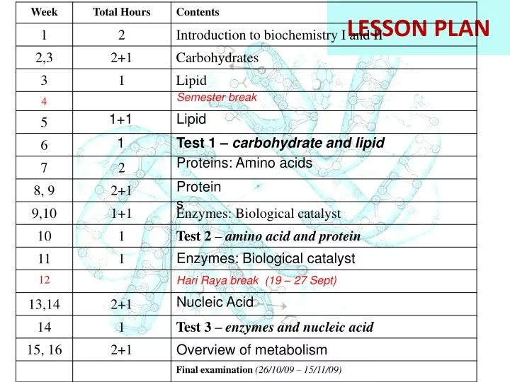
LESSON PLAN. Semester break. 1+1. Lipid. 1. Test 1 – carbohydrate and lipid. Proteins: Amino acids. Proteins. Enzymes: Biological catalyst. Hari Raya break (19 – 27 Sept). Nucleic Acid. Overview of metabolism. Protein: MONOMER – AMINO ACID. What is protein?.

E N D
LESSON PLAN Semester break 1+1 Lipid 1 Test 1 – carbohydrate and lipid Proteins: Amino acids Proteins Enzymes: Biological catalyst Hari Raya break (19 – 27 Sept) Nucleic Acid Overview of metabolism
What is protein? Proteinsarepolymers of amino acids. Primary Structure Secondary Structure Tertiary Structure Quaternary Structure
What is amino acid? Amino acid:a compound that contains both an amino group and a carboxyl groupattach to -carbon • -carbon also bound to side chain group, R • R gives identity to amino acid
Terminology • - carbon = the carbon that attach next to the carboxyl group • - amino group = amino group that attach to -carbon • Other type of amino group – eg. in Lysine, has • -amino group Lysine
Amino acid • All 20 are -amino acids 2. For 19 of the 20, the -amino group is primary; for proline, it is secondary amino acid -Amino acid has an amino group attached to the carbon (-carbon) adjacent to the carboxyl group
Generic amino acid at physiological pH amino acids exist as dipolar ionic species (have positive and negative charge on the same molecule) - zwitterion form Physiological pH Amino acid is an amphoteric molecule – act either as an acid or a base Amino acids as dipolar ions - carboxyl group carboxylate ion - amino group protonated amino acid
Enantiomer Mirror plane • The amino acids can exist in two enantiomeric forms (nonsuperimposable mirror image) forms – exceptional for glycine • Two steroisomers of amino acids are designated L- or D-. L – amino acid: abundant in nature, found in proteins, amino group on the left a carbon
Amino acid • Only the L - form of amino acids is commonly found in proteins. • Depending on the nature of the R group, amino acids are classified into four groups. 1. nonpolar 2. polar – neutral/uncharged side chain 3. acidic 4. basic Vs monosaccharide : D - form Polar, charged
Classification of amino acid • Nonpolar (9 amino acids) • Polar neutral/uncharged (6 amino acids) charged basic (3 amino acids) acidic (2 amino acids)
Classification of amino acids Simplest amino acid due to the R group = H No stereoisomer because the is achiral Nonpolar
Aliphatic cyclic structure – N is bonded to C2 atoms Amino group of become secondary amine – often called an imino acid Amino acids with nonpolar side chains - hydrophobic
Polar uncharged Amide bond – highly polar Phenol Thiol / sulfhydryl group – polar – under oxidizing condition, with other thiol groups to form disulfide bridges (-S-S-) – important in 3o structure
Polar charged Basic Aspartate Acidic Glutamate
Essential Amino acid • An essential amino acid or indispensable amino acid is an amino acid that cannot be synthesizedde novo bythe organism (usually referring tohumans), and therefore must be supplied in the diet. • vs non-essential amino acid
Ionization of Amino Acids • Remember, amino acids without charged groups on side chain exist in neutral solution as zwitterions with no net charge In acidic solution – as base (protonation) In basic solution – as acid (deprotonation)
Ionization of amino acids • At physiological pH, the carboxyl group of the amino acid is negatively charged and the amino group is positively charged. • Amino acids without charged side chains (Group 1 and 2) are zwitterions and have no net charge. (H3+N-HCR-COO- ). • A titration curve shows how the amine and carboxyl groups react with hydrogen ion.
Titration of amino acid • At low pH a nonacidic/nonbasic amino acid is protonated and has the structure H3N+HCRCOOH (amino acid in cationic form) • Increase of pH, dissociation of proton (H+) from –COOH group form H3N+HCRCOO- (amino acid in zwitterionic form) • At pK1, amount of cationic form = amount of zwitterionic form • Beyond pK1, additional base ions will results in all amino acids in cationic forms deprotonated to zwitterionic forms – all amino acids have no net charge pI = isoelectric point = pH at which the amino acid has no net charge/all amino acids are in zwitterionic form • Increase of pHbeyond pI, will cause the dissociation of H+ / deprotonation from H3N+resulting in formation of H2NHCRCOO- (anionic form) • Increase of pH, more dissociation of proton (H+) from –H3N+group, more amino acids in anionic form • At pK2, amount of zwitterionic form = amount of anionic form
Titration of Alanine • When an amino acid is titrated, the titration curve represents the reaction of each functional group with the hydroxide ion Anionic form pI (isoelectric point) = pH at which the amino acid has no net charge/ all amino acids are in zwitterionic form All amino acids are in the zwitterion form – at isoelectric point (pI) Cationic form
Titration of amino acid • pK1 and pK2 are proton dissociation constant from carboxyl group and amino group • From titration of amino acid, the pI can be calculated • The charge behavior of acidic and basic amino acids is more complex. – Group Polar/charged amino acid
Terminology • peptide: the name given to a short polymer of amino acids joined by peptide bonds; they are classified by the number of amino acids in the chain • dipeptide: a molecule containing two amino acids joined by a peptide bond • tripeptide: a molecule containing three amino acids joined by peptide bonds • polypeptide: a macromolecule containing many amino acids joined by peptide bonds • protein: a biological macromolecule of molecular weight 5000 g/mol or greater, consisting of one or more polypeptide chains Primary structure = one polypeptide
Peptide * * * * * Amino acid residue: a monomeric unit of amino acids
Primary structure Primary (1o) Structure = sequence of a chain of amino acids. Determines the final structure, eventually the properties of proteins
Peptide bond • The amino acids are linked through peptide bond • Peptide bond: the special name given to the amide bondbetween the -carboxyl group of one amino acid and the -amino group of another amino acid • peptide bond – covalent bond
Peptide bond: Feature 5 1 2 3 4 Free rotation COO- NH3+ Peptide bond – in trans configuration, acts as a rigid and planar unit. Has limited rotation around the peptide bond (C-N).
Secondary structure • The planar peptide group and free rotating bonds between C-N and C-C are important • Two types: -helix and -pleated sheet • 2o structure: involves the hydrogen-bonded arrangement of the backbone of the protein N O
Secondary structure: -helix • Structural features: • One polypeptide chain • Hydrogen bonds between the -CO and the –NH in the same polypeptide chain (intrachain) • The hydrogen bonds are parallel to the helix axis • Winding can be right- or left- handed (L- amino acid favor right-handed) H bond N O
Secondary structure: -pleated sheet • Structural features: • More than one polypeptide chain • Two types: antiparallel and parallel pleated sheet • Hydrogen bonds between the -CO and the –NH in the same polypeptide chain or with other polypeptide chain (interchain) • The hydrogen bonds are perpendicular to the direction of chain
Secondary structure: -pleated sheet • antiparallel pleated sheet = peptide chains are in the opposite directions • parallel pleated sheet = chains are in the same direction, the N- and C- terminal ends are aligned
Tertiary structure • Results from folding and packing of secondary structure • Bring together amino acid residues far apart, permitting interactions among their side chains Tertiary structure Is the three-dimensional arrangement of all atoms in protein molecule
Tertiary structure • Is the three-dimensional arrangement of all atoms in protein molecule • Involves non-covalent interaction and covalent bonds • Hydrogen bonds between the side chain • Hydrophobic interaction • Electrostatic interactions/attractions • Disulfide bonds – between the R group • Complexation with metal ions
Forces in 3˚ Structure • Noncovalent interactions, including • hydrogen bonding between polar side chains, e.g., Ser and Thr • hydrophobic interaction between nonpolar side chains, e.g., Val and Ile • electrostatic attraction between side chains of opposite charge, e.g., Lys and Glu • electrostatic repulsion between side chains of like charge, e.g., Lys and Arg, Glu and Asp • Covalent interactions: Disulfide (-S-S-) bonds between side chains of cysteines
Native conformation: three-dimensional shape of a protein with biological activity • Tertiary or quaternary structures
Quaternary structure • Final level of protein structure • Association of more than one polypeptide chain to form a complex • Subunit = individual parts of a large protein molecule = polypeptide chain • Quaternary structure is the result of noncovalent interactions between two or more protein chains. • Noncovalent interactions • electrostatics, • hydrogen bonds, • hydrophobic 2 3 4 1
Quaternary Structure • Oligomers are multisubunit proteins with all or some identical subunits. • The subunits are called protomers. • two subunits are called dimers • four subunits are called tetramers
Quaternary structure • If a change in structure on one chain causes changes in structure at another site, the protein is said to be allosteric. • Many enzymes exhibit allosteric control features. • Hemoglobin is a classic example of an allosteric protein. – oxygen = positive cooperativity • Has four subunits = tetramers • Overall structure 22 • Heme - Fe Structure of Hemoglobin
Classification of protein • Proteins are classified in two ways: • Shape • Composition
Fibrous Proteins • Fibrous proteins: contain polypeptide chains organized approximately parallel along a single axis. They • consist of long fibers or large sheets • tend to be mechanically strong • are insoluble in water and dilute salt solutions • play important structural roles in nature
Globular Proteins • Globular proteins: proteins which are folded to amore or less spherical shape • they tend to be soluble in water and salt solutions • most of their polar side chains are on the outside and interact with the aqueous environment by hydrogen bonding and ion-dipole interactions • most of their nonpolar side chains are buried inside • nearly all have substantial sections of -helix and -sheet
Holo- protein Proteins by Composition • Simple protein (apoprotein) Contain only amino acids ex. serum albumin and keratin • Conjugated protein • simple protein (apoprotein) • prostetic group (nonprotein) ex. Glycoproteins, lipoproteins, metaloproteins - hemoglobin
Denaturation • Definition – complete loss of organized structure in a protein, destroys the physiological function of the protein. • Definition – The unfolding of protein • Eg. During cooking of egg • Albumin (white egg) – denatured by heat and changes from a clear, colorless solution to a white coagulum • Often irreversible – denatured protein cannot returned to its native biological form – lost of biological function – why microbes die when boiling
Due to loss of 2o 4o of protein structure, but not 1o , the amide bond (peptide bond) is intact
Denaturation • Several ways to denature proteins • • Heat – in temp, vibrations within the molecule, the energy of these vibrations can disrupt the 3o • • pH – or pH, affect the charges of protein, the electrostatic interactions that normally stabilize the native conformation is reduced. • • Detergents (eg. SDS) - disrupt hydrophobic interactions, if the detergent is charged, this can also disrupt electrostatic interactions • • Reducing agents(eg. Urea) – will form stronger H bonds, stronger than within the protein. Also disrupt the hydrophobic interaction • • Heavy metal ions • Mechanical stress
Denaturation • Reversible denaturation – organic solvents (ethyl alcohol or acetone), urea, detergents and acid or base • Denaturants disrupt only noncovalent interactions not the covalent linkages of the primary structure • If removed, possible protein to unwound to native structure • eg. pH – addition of picric acid, protein (casein) precipitate addition of NaOH, the solution clear
Denaturation • -mercaptoethanol example of reversible denaturation. • -mercaptoethanolreduced the disulfide bridges of protein the unfolding of 3o structure, • the removal of -mercaptoethanol will cause the oxidation of SH group to form disulfide bridges again and the 3o structure is recovered.
Protein Functions