Download
1 / 76
780 likes | 824 Views
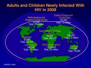
Gastrointestinal Manifestations of HIV-infected Children. Nuthapong Ukarapol, M.D. Division of Gastroenterology Department of Pediatrics Chiang Mai University. Introduction. GI involvement : one of most common complications in the HIV-infected patients

E N D
Gastrointestinal Manifestations of HIV-infected Children Nuthapong Ukarapol, M.D. Division of Gastroenterology Department of Pediatrics Chiang Mai University
Introduction • GI involvement : one of most common complications in the HIV-infected patients • Thea DM et al. reported that 37% of HIV-infected infants experienced diarrhea. (N Engl J Med 1993; 329:1696-702)
GI immunity and pathogenesis of diarrhea Altered GI immunity decreased IgA secretion decreased gastric secretion altered GI motility Predispose GI tract to Opportunistic infections Neoplasms
Common gastrointestinal symptoms & signs Diarrhea Abdominal pain Dysphagia and Odynophagia Gastrointestinal bleeding Weight loss and anorexia
HIV infection CD4-infected cells cross endothelium into the GI lamina propria HIV is uptaken by macrophage Decreased IgA Increased CD8 and lymphoid population in the lamina propria Dormant in the mesenteric lymph nodes Bacterial overgrowth Any opportunistic or non-opportunisticGI infection Increased enodtoxin Decreased CD4 population Accelerate viral replication T cell activation (CD4) Villous atrophy Crypt hypoplasia TNF, IFN anorexia Decreased lactase and disaccharidase activity Fat malabsorption Mucosal injury Villous atrophy Crypt hyperplasia malnutrition Malabsorption Malnutrition Diarrhea Pathogenesis of diarrhea in HIV-infected patients
Etiology of diarrhea in HIV-infected patients • Microsporidia, Isospora belli, and Cryptosporidium parvum infection : the first 3 most common pathogens detected in the HIV-infected patients with chronic diarrhea. (Kelly P Q J Med 1996; 89:813-7.)
Cryptosporidium Isospora belli Microspora
CMV MAI Cryptococcus neoformans Penicillium TB • Miller TL, et al. J Pediatr 1997; 130:766-73. • Rene E, et al. Dig Dis Sci 1989; 34:773-80.
Diagnosis: Diarrhea & HIV infection • Predictive factors for positive findings in EGD in the HIV-infected patients with diarrhea 1. AIDS stage 2. Serious bacterial infection 3. Many GI symptoms • Miller TL, et al. J Pediatr 1997; 130:766-73.
No gross abnormalities : NPV for normal histologic study:83% esophagus,79% stomach,65% duodenum Miller TL, et al. J Pediatr 1997; 130:766-73. Only 9.3 % of normal endoscopic findings were associated with histologic abnormalities Lim SG, et al. Gut 1993; 34:1429-32. Diagnosis: Diarrhea & HIV infection did not recommend routine surveillance biopsy in the patients who still have CD4 count over 200 /cumm, except in the patients with diarrhea recommended that tissue biopsies and cultures should be carried out while doing endoscopy
Diagnosis: HIV-infected children with diarrhea • Stool examination and cultures • Endoscopy • Ultrasound • CT abdomen Penicillium marneffei infection, Mycobacterium tuberculosis, and Mycobacterium avium-intracellulare
Diagnosis: Diarrhea & HIV infection • Randin DR. AJR 1991; 156:487-91.
Ultrastructure of Intestinal Biopsy in HIV-infected patients without identifiable pathogen • Irregular microvilli • joined bases microvilli • shortened and broadened microvilli • tubuloreticular inclusions in the endothelium cells • immune function disturbances and viral infections Fontana M, et al. J Pediatr Gastroenterol Nutr 1993; 17:255-9.
A 6 m/o HIV infected infant presented with chronic diarrhea. After extensive investigations, no specific causes could be identified. Irregular microvilli joined bases microvilli shortened and broadened microvilli
Rx: antiretroviral agents AZT & Lamivudine Outcomes: 1. Diarrhea stopped 2. Weaning off special formula 3. Gaining weight Restart medication stop medication start medication AIDS enteropathy
Abdominal pain & HIV infection • Other possibility : Penicillim marneffei mesenteric lymphadenitisUkarapol N, et al. J Med Assoc Thai 1998; 81:637-40.
Penicillim marneffei mesenteric lymphadenitisUkarapol N, et al. J Med Assoc Thai 1998; 81:637-40. • Report 3 cases of HIV-infected children with fever and abdominal pain: mimic acute abdomen • Physical signs of peritonitis were noted. • The first 2 patients were diagnosed as acute ruptured appendicitis and had an operation done. • The last patient was diagnosed as sepsis.
case 1 case 2 case 3 Investigations Hb/Hct 9.7/29.8 7.1/23 9.4/30 ( gm% /%) WBC(/x10-6 l) 3150 7400 4400 Plt(/ x10-6 l)14000073000 219000
case 1 case 2 case 3 Initial Tx exploratomy exploratomy Ceftriazone I.V. laparotomy laparotomy Operativenormal appendix normal appendix not done findings multiple and enlargement of matted mesenteric mesenteric nodes and paraaortic LN enlargement U/S abdomennot done multiple small* Matted enlarge round hypoechoic multiple LN around lesions at porta celiac artery and hepatis(LN) mesenteric vessels
Abdominal ultrasound History of acute abdomen with signs of peritonism Penicillium marneffei mesenteric lymphadenitis
case 1 case 2 case 3 mesenteric P. marneffei P. marneffei not done LN biopsy BM smear P. marneffei P. marneffei P. marneffei Skin smear not done P. marneffei not done BM cultureP. marneffei P. marneffei P. marneffei Hemoculture P. marneffei P. marneffei P. marneffei
Conclusion I. This report presented clinical manifestrations of P. marneffei infection which are different from previous reports including - abdominal pain - clinical signs which mimic peritonitis
II. We suggest things that might help to correct diagnosis. 1. history of HIV infection 2. skin lesions of P. marneffei infection 3. anemia, leukopenia and thrombocytopenia 4. abdoninal ultrasound 5. skin and bone marrow smear 6. blood and bone marrow culture 7. Endemic area of P. marneffei
Abominal pain yes Peritonitis no Prolonged fever Pancytopenia Skin lesion CBC, UA, stool exam&c/s serum amylase, lipase, LFTs plain abdomen yes no Blood culture Smear skin lesion Bone marrow exam&culture Ultrasound abdomen Explor Lower abdominal pain Upper abdominal pain EGD ERCP BE Colonoscopy P marneffei Diagnosis No diagnosis Diagnosis CT abdomen, liver biopsy
Dysphagia & Odynophagia in HIV-infected patients • Esophagitis, Esophageal ulcer • Candida albicans • Cytomegalovirus • Herpes simplex virus
Upper endoscopy: at the EG junction Erythema, Friability, Ulcer CMV Esophagitis Ulcer
Upper endoscopy: White plaques on the esophageal mucosa Candida Esophagitis
Dysphagia & Odynophagia in HIV-infected patients • Stoane JM, et al. Radiol Clin North Am 1996; 34:779-90.
GI bleeding & HIV infection • Non-infectious causes • Infectious causes e.g. salmanella, shigella, Campylobacter, E. coli, E. histolytica, CMV ileitis and CMV colitis • Penicillium marneffei • Mycobacterium tuberculosis, Mycobacterium avium-intracellulare • Diffuse infiltrative lymphocytosis syndrome in the stomach
Penicillium marneffei Colitis Penicillium marneffei : duodenal biopsy
Diffuse infiltrative lymphocytosis: in the stomach
Gastrointestinal cytomegalovirus disease in AIDS children Nuthapong Ukarapol1, Wattana Chartapisak1, Nirush Lertprasertsuk2, Lumduan Wongsawasdi1, Vinaisak Kattipattanapong3, Jesda Singhavejsakul3, Virat Sirisantana1 1 Department of Pediatrics, 2 Department of Pathology, 3 Department of Pediatric Surgery Faculty of Medicine, Chiang Mai University, Thailand
Patients & Methods • 1995-2001 • 8 patients with histologically confirmed gastrointestinal CMV infection were retrospectively reviewed.
Results: • 6 of 8 < 1 year old • median age 4.5 months (2 months-8 year 7 months)
Laboratories • 2 patients had a CD4 count done: with severe immunosuppression in 1 patient. • CD4 count (cells/µl) : 1080 (33%), 490 (16%) • 2 patients were diagnosed as CMV retinitis • 1 patient also had CMV pneumonitis • 1 patient was suspected having CMV hepatitis
Endoscopic findings: • 4 colonoscopy • 3 EGD • 1 flexible sigmoidoscopy Indications 1. Lower GI hemorrhage 2. Chronic diarrhea 3. Odynophagia
Endoscopic findings • mucosal edema • loss of normal vascular pattern • patchy erythema • friability • multiple ulcers Included