Download
1 / 89
940 likes | 1.46k Views
Coronary Heart Disease. NUR -224. Coronary Heart Disease. Affects 16 million people Causes more than 600,000 death annually Caused by impaired blood flow to the myocardium Accumulation of atherosclerotic plaque in the coronary arteries usual cause.

E N D
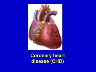
Coronary Heart Disease NUR -224
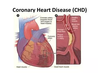
Coronary Heart Disease • Affects 16 million people • Causes more than 600,000 death annually • Caused by impaired blood flow to the myocardium • Accumulation of atherosclerotic plaque in the coronary arteries usual cause. • May be asymptomatic or may lead to angina pectoris, acute coronary care syndrome, myocardial infarction (MI/heart attack), dysrhythmias, heart failure and even sudden death
Coronary Atherosclerosis • Atherosclerosis is the abnormal accumulation of lipid deposits within arterial walls and lumen. • In coronary atherosclerosis, blockages and narrowing of the coronary vessels reduce blood flow to the myocardium.
Coronary Heart Disease Risk Factors • age (over 50) • heredity • smoking • obesity • high serum cholesterol levels • hypertension • diabetes mellitus
Angina Pectoris • Characterized by chest pain and usually precipitated by exercise and relieved by rest. • Myocardial oxygen needs are greater than partially occluded vessels can supply, myocardial cells become ischemic and shift to anaerobic metabolism • produces lactic acid that stimulates the nerve endings in the muscle pain • Pain subsides when oxygen supply meets myocardial demand.
Angina Pectoris • Chest pain reduced coronary blood flow temporary imbalance between myocardial blood supply and demand • This causes temporary/reversible myocardial ischemia. • More than 30 minutes of ischemia irreversibly damages myocardial cells (necrosis)
Types of Angina • Stable angina • Unstable angina • Silent Ischemia • Variant angina (Prinzmetal’s angina)
Stable Angina • Occurs with a predictable amount of activity or stress • Predictable and common • Occurs when the work of the heart is increased by physical exertion, exposure to cold, or by stress • Sensation may occur neck, jaw, shoulders • Lasts for few minutes – 5/15 minutes • Relieved by rest and/or nitrates
Unstable Angina • Occurs with increasing frequency, severity and duration • Pain is unpredictable and occurs with decreasing levels of activity or stress and may occur at rest. • Patients at risk for myocardial infarction • May not be relieved by NTG or rest
Variant Angina (Prinzmetal’s) • Atypical angina that occurs unpredictably • Caused by coronary artery spasm with/out atherosclerotic lesion • Often occurs at night
Silent Ischemia • Occurs in the absence of any subjective symptoms (asymptomatic) • Patient reports no pain • May occur with either activity or mental stress
Clinical Manifestations • Chest pain • Precipitated by an identifiable event. • Classic sequence • Women frequently present with atypical symptoms of angina
Angina Pain Manifestations • Chest Pain • Quality • Associated manifestations • Atypical manifestations • Precipitating factors • Relieving factors
Assessment/Diagnostic Findings • Past medical history/family history • Comprehensive description of chest pain • Presence of risk factors • Electrocardiography • Echocardiography • Cardiac stress testing • Cardiac Angiography
Medical Management • Focus on maintaining coronary blood flow and cardiac function • Measures to restore coronary blood flow (later) • Pharmacologic therapy * nitrates * beta blockers * calcium channel blockers * aspirin
Nitrates • SL NTG -> acute angina attacks (Buccal spray) • Acts within 1-2 minutes Long acting nitroglycerin • Used to prevent attacks not treat an acute attack. • Headache, hypotension, nausea, dizziness • NTG, Nitrostat, Nitro-bid
Nitrates • Nursing Responsibilities • Patient Teaching
B –Adrenergic Blockers • Decrease myocardial contractility, HR, BP which will reduce myocardial oxygen demand • Side effects bradycardia, hypotension, wheezing, GI complaints. • Contraindicated for patient with asthma • Metoprolol (Lopressor), Atenolol(Tenormin), Carvedilol (Coreg)
Beta Blockers Nursing Responsibilities Patient Teaching
Calcium Channel Blockers • Decrease the workload of the heart • Relax blood vessels decrease BP and increase coronary perfusion • Potent coronary vasodilator • Amlodipine (Norvasc), Diltiazem(Cardizem), Felodipine (Plendil) • Used to treat Variant Angina
Aspirin • Prevent platelet aggregation/thrombus formation • Reduces the incidence of MI • 80-325 mg of aspirin as soon as dx. is made • If patient is taking Tylenol – should continue to take aspirin • GI upset – H2 blocker, PPI
Nursing Diagnosis • Ineffective Cardiac Tissue Perfusion • Deficient Knowledge • Risk for Ineffective Therapeutic Regimen Management
Acute Coronary Syndrome • Condition of unstable cardiac ischemia • Includes unstable angina and acute myocardial ischemia c/out significant injury of myocardial tissue • Coronary blood flow is acutely reduced, but not fully occluded. Myocardial cells are injured by the acute ischemia that results. • Most people have stenosis of one or more coronary arteries.
Acute Coronary Syndrome Cardinal manifestation • Chest pain – substernal/epigastric • Dyspnea • Diaphoresis • Pallor • Cool skin • Tachycardia • Hypotension
Acute Coronary Care Syndrome Diagnosis • ECG • Cardiac Markers *Cardiac muscle troponins (sensitive indicators of myocardial damage) * Creatine Kinase (CK) & CK-MB (specific to myocardial muscle)
Medications • Reduce myocardial ischemia • Reduce risk for blood clotting • Nitrates • Beta blockers • Antiplatelet (po/IV)
Oral Antiplatelet -Medication • Aspirin • Clopidogrel (Plavix) • Ticlopidine (Ticlid) • Suppress platelet aggregation, prevents the development of thrombus. • Nursing responsibilities • Patient Education
IV Antiplatelet • Tirofiban (Aggrastat) • Eptifibatide (Integrilin) • IV antiplatelets more effective than oral administered meds but the risk of bleeding is greater
Heparin • Prevents the formation of new clots • Reducers the occurrence of MI • IV bolus/then continuous infusion • Infusion based on PTT – 2-2.5 times the normal PTT value (25-35 sec.) • LMWH – Lovenox/Fragmin • All increase the risk of bleeding : bleeding precautions
Revascularization Procedures • Several procedures may be used to restore blood flow and oxygen to ischemic tissue. Nonsurgical techniques: • percutaneous coronary revascularization • coronary atherectomy • intracoronary stents • coronary artery bypass graft (surgical procedure may be used)
Percutaneous Coronary Revascularization Is recommended for patients: • Fail medical management • Have left main coronary artery/three vessel disease • Are not candidates for PCI • Have failed PCI with ongoing chest pain
Percutaneous Coronary Intervention Goal: open the affected artery within 90 minutes of arrival to a facility Advantages: • Alternative to surgical intervention • Performed with local anesthesia • Patient is ambulatory 24 hours after the procedure • Hospital stay shorter • Patient can return to work
Percutaneous Coronary Revascularization • A balloon-tipped catheter is threaded over the guide wire • Balloon is inflated for about 30 sec.-2 minutes to compress the plaque against the arterial wall • The stent is then placed over a balloon catheter and expanded as the balloon is inflated • It remains in the artery when the balloon is removed.
Percutaneous Coronary Intervention Post procedure care: • Assess vital signs • Bedrest/ flat in bed • Affected leg straight • Pressure dressing applied • Monitor for bleeding/hematoma • Resume self-care activities/ambulation few hours after procedure
Atherectomy • Remove plaque from the identified lesion • Catheter shaves the plaque off vessels walls using a rotary cutting head - retaining the fragments in it compartment and removing them from the vessel. • Rotation catheter pulverize the plaque into particles small enough to pass through the coronary microcirculation.
CABG • Involves using a section of a vein /artery to create a connection (bypass) between the aorta and the coronary artery beyond construction. • This allows blood to perfuse the ischemic portion of the heart. • Internal mammary artery in the chest/saphenous vein from the leg are the vessels most commonly used.
CABG Surgery • Internal mammary artery – is commonly used. • Remains palliative treatment and not a cure. • Improves quality of life/patient outcomes • Postoperative complications/mortality increase as a function of age. • Women have a higher mortality rate than men
Patient teaching • Lifestyle changes and reduction of risk factors • Explore, recognize, and adapt behaviors to avoid to reduce the incidence of episodes of ischemia • Teaching regarding disease process • Cardiac rehabilitation • Stress reduction • When to seek emergency care
Myocardial Infarction • An area of the myocardium is permanently destroyed necrosis of the myocardial cells. If circulation to the affected myocardium is not promptly restored , the heart loses the ability to maintain effective cardiac output. • Life-threatening event • May lead to cardiogenic shock and death
Myocardial Infarction • Annually 785,000 experience their first MI • Majority of deaths from MI occur during the initial period after symptoms begin: 60% within the first hour 40% prior to hospitalization • Medical treatment and training in CPR are vital to decrease deaths due to MI.
Myocardial Infarction • The area of infarction develops over minute to hours. • Cellular ischemia affects conduction and myocardial contractility • Myocardial contractility decreases, increasing the risk for dysrhythmias, subsequently reducing cardiac output, B/P, and tissue perfusion
Myocardial Infarction • When a larger artery is compromised, collateral vessels connecting smaller arteries in the coronary system dilate to maintain blood flow to the cardiac muscle. • The degree of collateral circulation determines the extent of myocardial damage. • Occlusion of coronary artery without any collateral vessels massive tissue damage and death
Clinical Manifestations • Pain – sudden and usually not associated with activity • Women/older adults atypical chest pain, elevated BP & HR initially, then ↓’es • Nausea/vomiting • Fever • Dyspnea, shortness of breath • Anxiety , sense of impending doom